Case Reports
. 2021 May 7:2021:6625689.
doi: 10.1155/2021/6625689. eCollection 2021.

Renal Cell Carcinoma Metastasis to the Breast: A Rare Presentation

Affiliations
Case Reports

Renal Cell Carcinoma Metastasis to the Breast: A Rare Presentation

Heba O E Ali et al. Case Rep Radiol. .

Abstract

Worldwide breast malignancy is the most common cancer in women; however, metastases to the breast from extramammary malignancies are very rare and only a few sporadic cases are reported in the international literature. In this article, the authors report a case of a 73-year-old woman, who underwent nephrectomy for clear cell renal cell carcinoma and 3 years later presented with a breast metastasis from renal cell carcinoma (clear cell type).


Conflict of interest statement

The authors declare that they have no conflicts of interest.

Figures

Figure 1
Figure 1
Ultrasound showing malignant looking left renal mass with mixed solid and cystic component.
Figure 2
Figure 2
Ultrasound showing hypoechoic masses within the pancreas.
Figure 3
Figure 3
CT showing confirmed a large malignant looking left renal mass with central necrosis.
Figure 4
Figure 4
Follow-up CT of the abdomen showing liver deposits.
Figure 5
Figure 5
CT of the brain with contrast showing brain metastasis with surrounding edema.
Figure 6
Figure 6
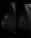
(a) Bilateral mammogram MLO view showing rounded density in the upper part of the right breast. (b) Mammogram in CC view showing a rounded right breast dense lesion.
Figure 7
Figure 7
New lesion as compared to previous mammogram.
Figure 8
Figure 8
US showing a hypoechoic solid mass lesion in the right breast at 1 o'clock.
Figure 9
Figure 9
The standard H&E-stained section showing cells with small nuclei and abundant clear cytoplasm typical of clear cell RCC.
Figure 10
Figure 10
Image shows positive immunohistochemical cell nucleus staining with PAX8.
Figure 11
Figure 11
Image shows positive cell membrane staining with carbonic anhydrase.

Similar articles

Cited by

References

    1. Padala S. A., Barsouk A., Thandra K. C., et al. Epidemiology of renal cell carcinoma. World Journal of Oncology. 2020;11(3):79–87. doi: 10.14740/wjon1279. - DOI - PMC - PubMed
    1. Howlander N., Noone A. M., Krapcho M. SEER cancer statistics review 1975-2016. National Cancer Institute; 2019.
    1. Campbell S. C., Lane B. R. Malignant renal tumors. Campbell-Walsh Urol; 2012.
    1. Wahner-Roedler D. L., Sebo T. J. Renal cell carcinoma: diagnosis based on metastatic manifestations. Mayo Clinic Proceedings. 1997;72(10):935–941. doi: 10.4065/72.10.935. - DOI - PubMed
    1. Tarraza H. M., Jr., Meltzer S. E., DeCain M., Jones M. A. Vaginal metastases from renal cell carcinoma: report of four cases and review of the literature. European Journal of Gynaecological Oncology. 1998;19(1):14–18. - PubMed

Publication types

LinkOut - more resources