

Characterization of DDX4 Gene Expression in Human Cases with Non-Obstructive Azoospermia and in Sterile and Fertile Mice
- PMID: 34041004
- PMCID: PMC8143011
- DOI: 10.18502/jri.v22i2.5793
Characterization of DDX4 Gene Expression in Human Cases with Non-Obstructive Azoospermia and in Sterile and Fertile Mice
Abstract
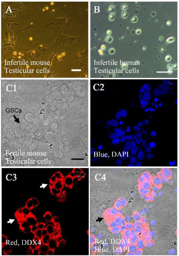
Background: In mammals, spermatogenesis is the main process for male fertility that is initiated by spermatogonial stem cells (SSCs) proliferation. SSCs are unipotent progenitor cells accountable for transferring the genetic information to the following generation by differentiating to haploid cells during spermato-and spermiogenesis. DEAD-box helicase 4 (DDX4) is a specific germ cell marker and its expression pattern is localized to, spermatocytes, and spermatids. The expression in the SSCs on the basement membrane of the seminiferous tubules is low.
Methods: Immunohistochemistry (IHC) and Fluidigm reverse transcriptase-polymerase chain reaction (RT-PCR) were used to analyze the expression of DDX4 in testis tissue of fertile and sterile mice and human cases with non-obstructive azoospermia.
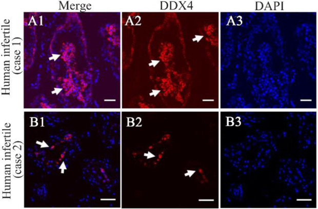
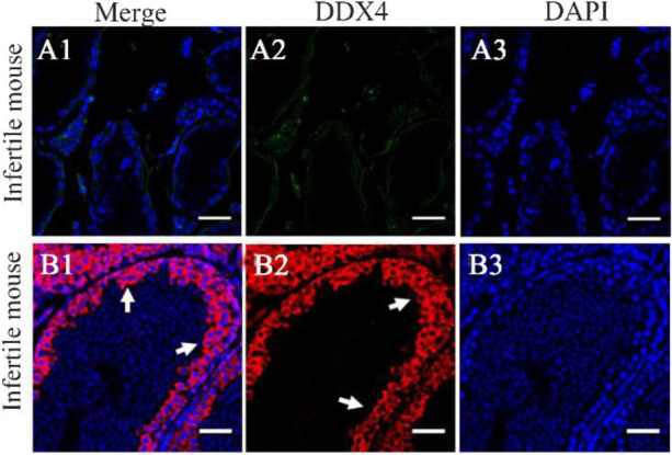
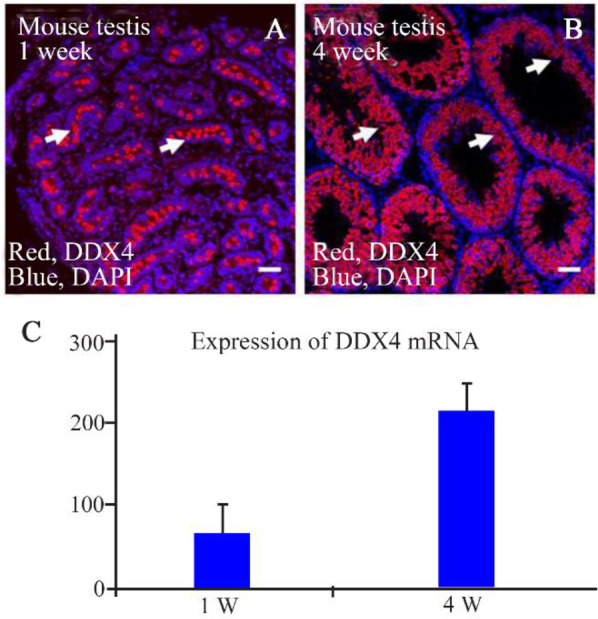
Results: Our immunohistochemical findings of fertile and busulfan-treated mice showed expression of DDX4 in the basal and luminal compartment of seminiferous tubules of fertile mice whereas no expression was detected in busulfan-treated mice. The immunohistochemical analysis of two human cases with different levels of non-obstructive azoospermia revealed more luminal DDX4 positive cells.
Conclusion: Our findings indicate that DDX4 might be a valuable germ cell marker for analyzing the pathology of germ cell tumors and infertility as global urological problems.
Keywords: DDX4 protein; Seminiferous tubule; Spermatogonial Stem Cell; Testicles.
Copyright© 2021, Avicenna Research Institute.
Conflict of interest statement
Conflict of Interest The authors declare that they have no competing interests.
Figures
References
-
- Huang P, Wang T. Spermatogonial stem cell and TGF-\b {eta} involved regulation of proliferation and differentiation. arXiv preprint arXiv:1706. 03892. 2017. June 13.
-
- Meinhardt A, Wang M, Schulz C, Bhushan S. Microenvironmental signals govern the cellular identity of testicular macrophages. J leukoc Biol. 2018;104(4):757–66. - PubMed
-
- Wang F, Han D. Sertoli cell phagocytosis: an essential event for spermatogenesis. In: Wu W, Ziglioli F, Maestroni U, editors. Male reproductive health. UK: IntechOpen; 2019. p. 69–84.
LinkOut - more resources
Full Text Sources
Other Literature Sources