. 2021 May 10:11:646546.
doi: 10.3389/fcimb.2021.646546. eCollection 2021.

Exosomes Derived From Alveolar Epithelial Cells Promote Alveolar Macrophage Activation Mediated by miR-92a-3p in Sepsis-Induced Acute Lung Injury

Affiliations

Exosomes Derived From Alveolar Epithelial Cells Promote Alveolar Macrophage Activation Mediated by miR-92a-3p in Sepsis-Induced Acute Lung Injury

Fen Liu et al. Front Cell Infect Microbiol. .

Abstract

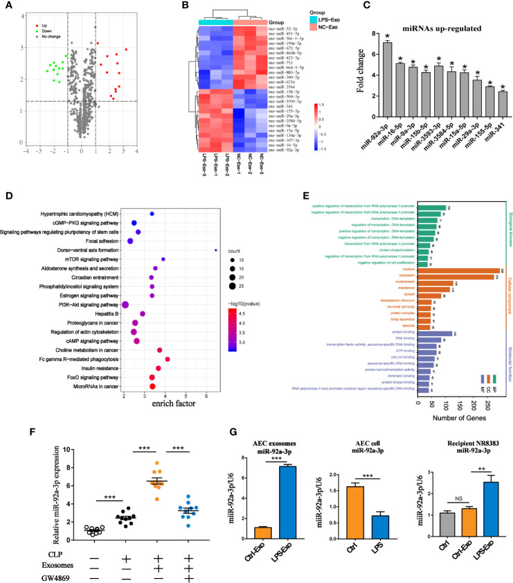
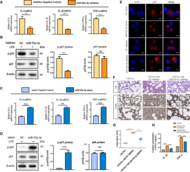
Acute lung injury (ALI) induced by sepsis is characterized by disruption of the epithelial barrier and activation of alveolar macrophages (AMs), which leads to uncontrolled pulmonary inflammation. However, effective treatments for ALI are unavailable. The exact mechanism by which the initial mediator of alveolar epithelial cells (AECs) induces inflammation remains elusive. Here we investigated the roles of AEC-derived exosomes in AM activation and sepsis-induced ALI in vivo and in vitro. Cecal ligation and puncture (CLP) was utilized to establish septic lung injury model in rats. The effect of exosomal inhibition by intratracheal GW4869 administration on lung injury was investigated. To assess the effects of AEC-derived exosomes on ALI, we treated the rat alveolar epithelial cell line RLE-6TN with LPS to induce cell damage. Exosomes from conditioned medium of LPS-treated AECs (LPS-Exos) were isolated by ultracentrifugation. The miRNAs in LPS-Exos were screened by miRNA expression profile analysis. The effects of miR-92a-3p on the function of AMs were studied. We found that intratracheal GW4869 administration ameliorated lung injury following CLP-induced ALI. LPS-Exos were taken up by AMs and activated these cells. Consistently, administration of LPS-Exos in rats significantly aggravated pulmonary inflammation and alveolar permeability. Moreover, miR-92a-3p was enriched in LPS-Exos and could be delivered to AMs. Inhibition of miR-92a-3p in AECs diminished the proinflammatory effects of LPS-Exos in vivo and in vitro. Mechanistically, miR-92a-3p activates AMs along with pulmonary inflammation. This process results in activation of the NF-κB pathway and downregulation of PTEN expression, which was confirmed by a luciferase reporter assay. In conclusion, AEC-derived exosomes activate AMs and induce pulmonary inflammation mediated by miR-92a-3p in ALI. The present findings revealed a previously unidentified role of exosomal miR-92a-3p in mediating the crosstalk between injured AEC and AMs. miR-92a-3p in AEC exosomes might represent a novel diagnostic biomarker for ALI, which may lead to a new therapeutic approach.

Keywords: acute lung injury; alveolar epithelial cells; alveolar macrophage; exosomes; miR-92a-3p; sepsis.


Conflict of interest statement

The authors declare that the research was conducted in the absence of any commercial or financial relationships that could be construed as a potential conflict of interest.

Figures

Figure 1
Figure 1
Inhibiting the secretion of intrapulmonary exosomes alleviates septic lung injury. Rats were treated with GW4869 (2.5 μg/g) through airway instillation. CLP procedures were conducted one hour post-treatment, and sham operated rats were used as negative controls. (A) Representative electron micrograph of exosomes purified from the BALF (indicated by white arrows). Scale bars, 200 nm. (B) Western blot assessment of exosomes showing the presence of CD63 and CD9 in BALF-derived exosomes. (C) Quantification of exosomal protein concentration in the BALF of each group of rats. (D) Histopathological examination of lung tissue by HE staining, Scale bars,50 µm. (E) Scoring the pathology of lung tissue damage using the Smith scoring method. (F–H) Changes in lung tissue oedema of rats under different treatments. (F) Evans blue content in lung tissue. (G) The wet/dry weight ratio in lung tissue. (H) The ratio of protein concentration in BALF to plasma. (I) The expression levels of IL-1β and TNF-α in BALF and plasma. (J, K) Survival rate of rats in each group within 7 days and the changes in sepsis score of each group. The Kaplan-Meier method combined with the log-rank test was used to compare multiple populations. Compared with the sham group, *P<0.05; compared with the CLP group, #P<0.05; n=10 for the sham group, n=20 for the other groups. (L) Western blot assessment of the expression of epithelial cell-specific marker proteins SP-B and cytokeratin in exosomes derived from BALF and quantitative analysis of the protein grey value. Data are presented as the mean ± S.E.M. **P<0.001, ***P<0.001 compared between two groups. n=6 per group.
Figure 2
Figure 2
Exosomes derived from LPS-treated AECs induce acute lung injury. (A–C) Characterization of exosomes isolated from the supernatant of cultured AECs. (A) Electron micrograph of exosomes. Scale bar, 100 nm. (B) Exosome size distribution was measured by Nano Sight tracking analysis. (C) Alix, CD63 and CD9 protein expression in exosomes was quantified by western blot loaded with equal amounts of protein (40 μg). (D–G) Exosomes (2 mg/kg) were intratracheally transferred to SD rats through intratracheal, and analysis was performed 24 h after treatment (n = 6 for each group). (D) Lung histological changes and lung injury scores were assessed with HE staining. Immunohistochemical detection of CD68 positive macrophage infiltration in the lung. The numbers of CD68 positive loci were quantified with ImageJ software. Scale bar, 50 μm and 25 μm. (E, F) ELISAs showed the expression of proinflammatory cytokines (IL-1β and TNF-α) in the BALF. (G) LPS-Exos labelled with PKH-67 fluorescent dye were administered to rats. Immunofluorescence images showing the colocalization between exosomes (green) and CD68- labelled AMs (red) in the lung. Nuclei were counterstained with DAPI (blue). Data are presented as the mean ± S.E.M. ***P < 0.001 compared between two groups. NS, no significant difference. n=6 per group.
Figure 3
Figure 3
Internalized LPS-treated AECs promote AM activation. (A) PKH-67 labelled AEC exosomes were cocultured with recipient AMs for 12 h. Relative fluorescence intensity was quantified to identify the internalization of exosomes. Scale bar, 30μm. (B) Proinflammatory cytokine IL-1β, TNF-α and IL-6 levels in the supernatant of the AMs treated with AEC exosomes for 12 h. (C, D) Representative western blot and quantitative analysis of p-p65 and p65 in recipient AMs treated with AEC-derived exosomes with or without LPS stimulation. (E) Representative images of the nuclear translocation of p65, as observed by immunofluorescence assays. Immunofluorescence staining of p65 (red) and nuclei (blue) at 12 h following AMs were treated with LPS-Exos or Ctrl-Exos. Scale bar, 5 μm. Data are presented as the mean ± S.E.M. **P<0.01, ***P<0.001 compared between two groups. NS, no significant difference. n=3 per group.
Figure 4
Figure 4
miRNA expression profile of LPS-treated AEC exosomes. (A) The volcano plot of differentially expressed miRNAs between the LPS-Exo and AEC-Exo groups (fold changes ≥2 and P<0.05), compared with the Ctrl-Exo group. The miRNAs with upregulated expression are represented by red dots, those with downregulated expression arerepresented by green dots and those with non-significantly changed expression are shown as grey dots. The vertical lines correspond to upregulation or downregulation by 2-fold, and the horizontal line represents the P value of 0.05. (B) The heatmap shows the differentially expressed miRNAs. (C) Validation of the expression levels of the top 10 upregulated miRNAs in LPS-Exos. Ctrl-Exos were used as a negative control. (D, E) Kyoto Encyclopedia of Genes and Genomes (KEGG) analysis and Gene Ontology (GO) were performed on the predicted target genes of miR-92a-3p. Ctrl-Exos and LPS-Exos represent exosomes isolated from the control and LPS-treated AECs, respectively. (F) The expression of miR-92a-3p in BALF or BALF-derived exosomes isolated from the septic rats. Rats were treated with GW4869 (2.5 µg/g) through airway instillation. An hour post-treatment, CLP procedures were conducted. Sham operated rats were used as negative controls. n=10 for each group. (G) Expression of miR-92a-3p in AEC derived exosomes, AECs and recipient AMs treated with AEC derived exosomes. Data are presented as the mean ± S.E.M. *P<0.05, **P<0.01, ***P<0.001 compared Brtween two groups. NS, no significance. n=3 per group.
Figure 5
Figure 5
AEC exosome-derived miR-92a-3p promotes alveolar macrophage inflammation in vitro and in vivo. (A, B) Inflammatory cytokine expression and the NF-κB signalling pathway in macrophages treated with exosomes from LPS-induced AECs transfected with miR-92a-3p inhibitor or the corresponding NC. (C, D) Inflammatory cytokine expression and the NF-κB signalling pathway in macrophages treated with exosomes from AECs transfected with miR-92a-3p mimic or the corresponding NC. (E) Representative images of the nuclear translocation of p65, as observed by immunofluorescence assays, Scale bar, 5 μm. Data are presented as the mean ± S.E.M.**P<0.01, ***P<0.001 compared between two groups. NS, no significant difference, n=3 per group. For the in vivo experiment, AECs were cultured and treated with PBS or LPS for 24 h after transfection with NC or miR-92a-3p inhibitor. Exosomes were transferred to rats through intratracheal instillation. Rats were sacrificed 24 h after the treatment (n = 6 for each group) (F) Histologic (HE staining) changes and CD68 positive macrophage infiltration in the lung tissue. (G) The numbers of CD68 positive loci were quantified. (H) ELISAs showed the concentration of intrapulmonary inflammatory cytokines (IL-1β, TNF-α) in the BALF. Data are presented as the mean ± S.E.M. ***P<0.001 compared between two groups. n=6 per group.
Figure 6
Figure 6
Exosomal miR-92a-3p activates alveolar macrophages via PTEN. (A) Conservation of the miR-92a-3p target sequence in the PTEN 3’UTR among different species and conservation of the miR-92a-3p sequence among different species. (B) The dual-luciferase reporter assay was performed in 293T cells. Cells were cotransfected with the wild-type or mutant PTEN 3’UTR luciferase reporter plasmids, as well as miR-92a-3p mimic or NC mimic. (C) mRNA and protein expression of PTEN in recipient alveolar macrophages treated with AEC-Exo were examined by RT-PCR and western blot analysis. (D) The protein levels of PTEN were detected using western blot analysis after transfection with miR-92a-3p inhibitor or miR-92a-3p mimic. β-actin was used as an internal control. (E) Immunoblot of lysates from AMs overexpressing PTEN in the presence or absence of LPS-Exos. The rescue of PTEN expression dramatically inhibited LPS-Exo induced p65 phosphorylation and tended to decrease these levels in response to LPS-Exos. β-actin was used as an internal control. Data are presented as the mean ± S.E.M. **P<0.01, ***P<0.001 compared between two groups. NS, no significant difference, n=3 per group.
Figure 7
Figure 7
A schematic diagram showing the role of exosomal miR-92a-3p from LPS-induced AECs in inducing AM activation in septic lung injury. LPS stimulation induces AECs to release an increased number of exosomes, which are enriched with miR-92a-3p. The AEC-derived exosomes are taken up by AMs. miR-92a-3p induced degradation of the PTEN mRNA in AMs, resulting in the downregulation of the PTEN protein level and subsequent activation of the NF-κB signalling pathway. The nuclear translocation of p65 further promotes the release of proinflammatory factors in AMs.

Similar articles

Cited by

References

    1. Arora S., Dev K., Agarwal B., Das P., Syed M. A. (2018). Macrophages: Their Role, Activation and Polarization in Pulmonary Diseases. Immunobiology 223, 383–396. 10.1016/j.imbio.2017.11.001 - DOI - PMC - PubMed
    1. Bissonnette E. Y., Lauzon-Joset J. F., Debley J. S., Ziegler S. F. (2020). Cross-Talk Between Alveolar Macrophages and Lung Epithelial Cells is Essential to Maintain Lung Homeostasis. Front. Immunol. 11, 583042. 10.3389/fimmu.2020.583042 - DOI - PMC - PubMed
    1. Chen X. H., Yin Y. J., Zhang J. X. (2011). Sepsis and Immune Response. World J. Emerg. Med. 2, 88–92. 10.5847/wjem.j.1920-8642.2011.02.002 - DOI - PMC - PubMed
    1. Chen T., Yu Q., Xin L., Guo L. (2020). Circular RNA circC3P1 Restrains Kidney Cancer Cell Activity by Regulating miR-21/PTEN Axis and Inactivating PI3K/AKT and NF- kB Pathways. J. Cell Physiol. 235, 4001–4010. 10.1002/jcp.29296 - DOI - PubMed
    1. Fein A. M., Calalang-Colucci M. G. (2000). Acute Lung Injury and Acute Respiratory Distress Syndrome in Sepsis and Septic Shock. Crit. Care Clin. 16, 289–317. 10.1016/S0749-0704(05)70111-1 - DOI - PubMed

Publication types