

Age-Dependent Remodeling in Infrapatellar Fat Pad Adipocytes and Extracellular Matrix: A Comparative Study
- PMID: 34041253
- PMCID: PMC8141643
- DOI: 10.3389/fmed.2021.661403
Age-Dependent Remodeling in Infrapatellar Fat Pad Adipocytes and Extracellular Matrix: A Comparative Study
Abstract
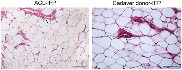
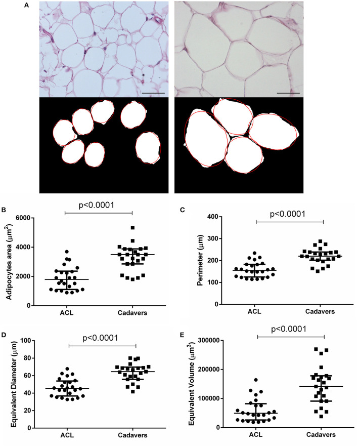
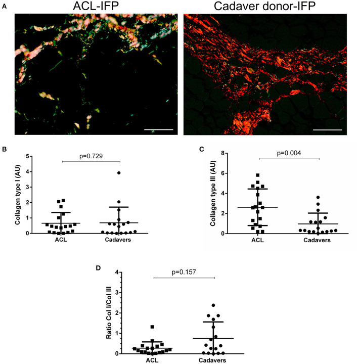
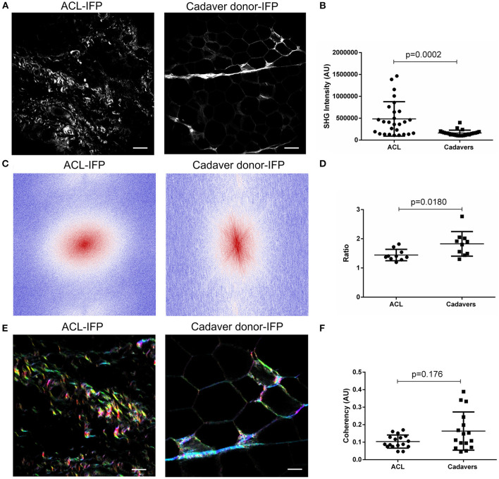
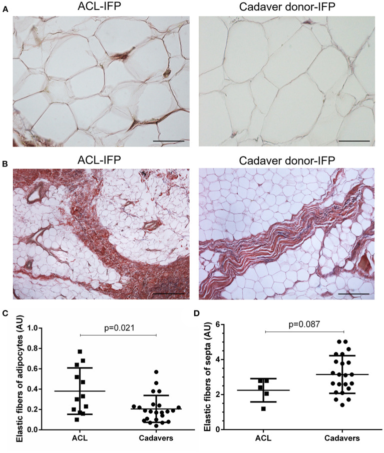
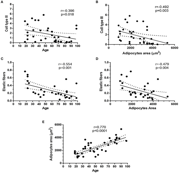
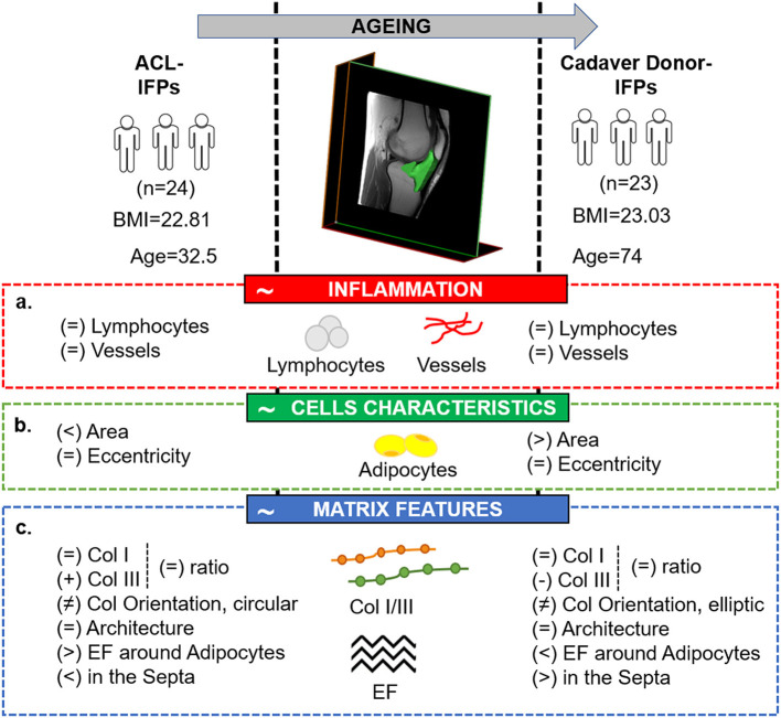
The infrapatellar fat pad (IFP) is actively involved in knee osteoarthritis (OA). However, a proper description of which developmental modifications occur in the IFP along with age and in absence of joint pathological conditions, is required to adequately describe its actual contribution in OA pathophysiology. Here, two IFP sources were compared: (a) IFP from healthy young patients undergoing anterior-cruciate ligament (ACL) reconstruction for ACL rupture (n = 24); (b) IFP from elderly cadaver donors (n = 23). After histopathological score assignment to confirm the absence of inflammatory features (i.e., inflammatory infiltrate and increased vascularity), the adipocytes morphology was determined; moreover, extracellular matrix proteins were studied through histology and Second Harmonic Generation approach, to determine collagens content and orientation by Fast Fourier Transform and OrientationJ. The two groups were matched for body mass index. No inflammatory signs were observed, while higher area, perimeter, and equivalent diameter and volume were detected for the adipocytes in the elderly group. Collagen III displayed higher values in the young group and a lower total collagen deposition with aging was identified. However, collagen I/III ratio and the global architecture of the samples were not affected. A higher content in elastic fibers was observed around the adipocytes for the ACL-IFPs and in the septa cadaver donor-IFPs, respectively. Age affects the characteristics of the IFP tissue also in absence of a pathological condition. Variable mechanical stimulation, depending on age-related different mobility, could be speculated to exert a role in tissue remodeling.
Keywords: aging; anterior-cruciate ligament rupture; cadaver donors; extracellular matrix; infrapatellar fat pad; osteoarthritis.
Copyright © 2021 Stocco, Belluzzi, Contran, Boscolo-Berto, Picardi, Guidolin, Fontanella, Olivotto, Filardo, Borile, Romanato, Ramonda, Ruggieri, Favero, Porzionato, De Caro and Macchi.
Conflict of interest statement
The authors declare that the research was conducted in the absence of any commercial or financial relationships that could be construed as a potential conflict of interest.
Figures
Similar articles
-
Exploring Anatomo-Morphometric Characteristics of Infrapatellar, Suprapatellar Fat Pad, and Knee Ligaments in Osteoarthritis Compared to Post-Traumatic Lesions.Biomedicines. 2022 Jun 9;10(6):1369. doi: 10.3390/biomedicines10061369. Biomedicines. 2022. PMID: 35740391 Free PMC article.
-
Infrapatellar Fat Pad Gene Expression and Protein Production in Patients with and without Osteoarthritis.Int J Mol Sci. 2020 Aug 21;21(17):6016. doi: 10.3390/ijms21176016. Int J Mol Sci. 2020. PMID: 32825633 Free PMC article.
-
The suprapatellar fat pad: A histotopographic comparative study.J Anat. 2024 Apr;244(4):639-653. doi: 10.1111/joa.13984. Epub 2023 Nov 29. J Anat. 2024. PMID: 38030148 Free PMC article.
-
Evaluation and treatment of disorders of the infrapatellar fat pad.Sports Med. 2012 Jan 1;42(1):51-67. doi: 10.2165/11595680-000000000-00000. Sports Med. 2012. PMID: 22149697 Review.
-
Inflammation of the infrapatellar fat pad.Joint Bone Spine. 2016 Jul;83(4):389-93. doi: 10.1016/j.jbspin.2016.02.016. Epub 2016 Apr 7. Joint Bone Spine. 2016. PMID: 27068617 Review.
Cited by
-
Exploring Anatomo-Morphometric Characteristics of Infrapatellar, Suprapatellar Fat Pad, and Knee Ligaments in Osteoarthritis Compared to Post-Traumatic Lesions.Biomedicines. 2022 Jun 9;10(6):1369. doi: 10.3390/biomedicines10061369. Biomedicines. 2022. PMID: 35740391 Free PMC article.
-
Quantification of infrapatellar fat pad fibrosis using magnetic resonance imaging-derived proton density fat fraction: a pathology-controlled study.Quant Imaging Med Surg. 2025 Apr 1;15(4):2694-2706. doi: 10.21037/qims-24-2021. Epub 2025 Mar 18. Quant Imaging Med Surg. 2025. PMID: 40235820 Free PMC article.
-
Impaired Remodeling of White Adipose Tissue in Obesity and Aging: From Defective Adipogenesis to Adipose Organ Dysfunction.Cells. 2024 Apr 30;13(9):763. doi: 10.3390/cells13090763. Cells. 2024. PMID: 38727299 Free PMC article. Review.
-
Quantitative MRI reveals infrapatellar fat pad changes after running a marathon.PeerJ. 2025 Mar 20;13:e19123. doi: 10.7717/peerj.19123. eCollection 2025. PeerJ. 2025. PMID: 40124601 Free PMC article.
-
Hydrostatic Pressure Enhances Chondrogenic Differentiation of Mesenchymal Stem Cells in Silk Fibroin-Based 3D Bioprinted Hydrogels.Biomacromolecules. 2025 Jun 9;26(6):3432-3445. doi: 10.1021/acs.biomac.5c00048. Epub 2025 May 20. Biomacromolecules. 2025. PMID: 40393648 Free PMC article.
References
-
- United Nations Secretariat , E.S.A. World Population Prospects: The 2017 Revision. Key Findings and Advance Tables. New York, NY: United Nations; (2017).
LinkOut - more resources
Full Text Sources
Other Literature Sources