. 2021 May 10:8:684934.
doi: 10.3389/fmolb.2021.684934. eCollection 2021.

Yin and Yang in Post-Translational Modifications of Human D-Amino Acid Oxidase

Affiliations

Yin and Yang in Post-Translational Modifications of Human D-Amino Acid Oxidase

Silvia Sacchi et al. Front Mol Biosci. .

Abstract

In the central nervous system, the flavoprotein D-amino acid oxidase is responsible for catabolizing D-serine, the main endogenous coagonist of N-methyl-D-aspartate receptor. Dysregulation of D-serine brain levels in humans has been associated with neurodegenerative and psychiatric disorders. This D-amino acid is synthesized by the enzyme serine racemase, starting from the corresponding L-enantiomer, and degraded by both serine racemase (via an elimination reaction) and the flavoenzyme D-amino acid oxidase. To shed light on the role of human D-amino acid oxidase (hDAAO) in D-serine metabolism, the structural/functional relationships of this enzyme have been investigated in depth and several strategies aimed at controlling the enzymatic activity have been identified. Here, we focused on the effect of post-translational modifications: by using a combination of structural analyses, biochemical methods, and cellular studies, we investigated whether hDAAO is subjected to nitrosylation, sulfhydration, and phosphorylation. hDAAO is S-nitrosylated and this negatively affects its activity. In contrast, the hydrogen sulfide donor NaHS seems to alter the enzyme conformation, stabilizing a species with higher affinity for the flavin adenine dinucleotide cofactor and thus positively affecting enzymatic activity. Moreover, hDAAO is phosphorylated in cerebellum; however, the protein kinase involved is still unknown. Taken together, these findings indicate that D-serine levels can be also modulated by post-translational modifications of hDAAO as also known for the D-serine synthetic enzyme serine racemase.

Keywords: D-serine; biochemical properties; flavoprotein; nitrosylation; phosphorylation; sulfhydration.


Conflict of interest statement

The authors declare that the research was conducted in the absence of any commercial or financial relationships that could be construed as a potential conflict of interest.

Figures

FIGURE 1
FIGURE 1
Cysteine residues in hDAAO. (A) Weblogo representation of conserved residues identified by the alignment of DAAO sequences from Homo sapiens, Mus musculus, Rattus norvegicus, Sus scrofa, Bos taurus, Cavia porcellus, and Macaca fascicularis. The x-axis represents amino acid positions (the annotated numbering refers to the human enzyme). The height of symbols is proportional to the degree of conservation of single residues. Panels represent sequence stretches of 20 residues containing cysteine residues, shown in red. Five cysteine residues appear highly conserved in mammalian DAAOs. Figure prepared using WebLogo (https://weblogo.berkeley.edu/logo.cgi). (B) Solvent-accessible surface (SAS) as calculated for the hDAAO dimer (pdb code 2E49). Cysteine residues are represented as sphere (carbon atoms in cyan, sulfur atoms in yellow), the FAD cofactor is represented as sticks (yellow), and the backbone is shown as cartoon. Protein surface is colored by proximity of cysteine residues (cyan = cysteine residues located within 5 Å from the surface; orange = cysteine residues located at a distance higher than 15 Å from the surface). SAS has been calculated based on a solvent radius of 1.4 Å. Two of the conserved cysteine residues are likely accessible to modifications. (C) Details showing the amino acidic environment of selected cysteine residues, represented as spheres (carbon atoms in cyan, sulfur atoms in yellow, nitrogen atoms in blue, and oxygen atoms in red). The surrounding residues are depicted as sticks; the van der Waals surface is shown. Hydrophobic residues are colored in orange, while charged residues are colored by element (carbon atoms in magenta, nitrogen atoms in blue, and oxygen atoms in red). Illustrations prepared with “The Protein Imager online molecular viewer” (https://3dproteinimaging.com/protein-imager/).
FIGURE 2
FIGURE 2
S-Nitrosylation of hDAAO induces partial inactivation. (A) The treatment of U87 cells ectopically expressing hDAAO with the NO donors NOR3 and NOC7 reduced the activity of the flavoenzyme. hDAAO-expressing cells were treated with 50 µM NOC7 (red squares), 50 µM NOR3 (blue squares), or with an equal amount of DMSO (CTRL, black circles) for 2 h. Cells transfected with the pcDNA3 empty vector and treated with the same amounts of the NO donors (blue open circles) or DMSO (black open square) were used as further controls. In treated and control cells hDAAO activity was detected in cell lysates by the Amplex UltraRed assay and reported as fluorescence emission at 590 nm over time. Values are reported as mean ± SEM, n = 5. (B) hDAAO is nitrosylated in vitro. The nonreducing SDS-PAGE analysis of recombinant hDAAO (7.5 and 15 µg) following in vitro S-nitrosylation and the fluorescence switch assay is shown. Mixtures in which the NO donor GSNO was replaced with GSH were analyzed as negative controls whereas the positive controls (CTRL) were represented by protein samples in which all cysteine residues were labeled by the Alexa Fluor 350 C5 Maleimide reagent (by omitting the MMTS blocking step during the fluorescence switch assay). Image acquisition was performed by a normal (top panel) and a CCD (middle panel) camera upon excitation of the fluorescent probe. Proteins were also stained with Coomassie blue (bottom panel). (C, D) Nitrosylation slightly alters hDAAO enzymatic stability without affecting the binding of the FAD cofactor. (C) Effect of the presence of GSNO on hDAAO activity, as determined by the oxygen consumption activity assay. In vitro nitrosylation mixtures and related controls were set up in the absence or presence of exogenous FAD (2.5 and 25 µM final concentration). Residual activity values (100% represents the initial activity of the unreacted enzyme) are reported as mean ± SEM, n = 5, ***p < 0.005, unpaired t-test. (D) Analysis of FAD binding to hDAAO apoprotein in the absence and presence of 50 or 500 µM of the NO donor GSNO, assessed as protein fluorescence quenching. Values are expressed as percentage of the total change in a single experiment (the determined Kd values are the average of three independent experiments). Left panel shows the interpolation of the experimental data corresponding to the first saturation phase of the protein fluorescence change (up to 2 µM of cofactor concentration); right panel reports the same analysis for the second phase of saturation. On the y-axis, 100% correspond to the change in fluorescence emission between the hDAAO apoprotein free form and the fully FAD-complexed one. Measurements were performed at 15°C. Black squares and continuous lines: control mixture in the absence of GSNO; blue circles and staggered lines: mixture containing 50 µM GSNO; red triangles and dotted lines: mixture containing 500 µM GSNO.
FIGURE 3
FIGURE 3
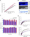
Effect of S-sulfhydration on hDAAO properties. (A) Treatment of U87 transfected cells expressing hDAAO with the hydrogen sulfide donor NaHS increases the activity of the flavoenzyme. Cells were treated with 50 µM NaHS (red squares) or with an equal amount of H2O (CTRL, black circles) for 30 min. Cells transfected with the pcDNA3 empty vector and treated with the same amount of the H2S donor (open circles) were used as further controls. In treated and control cells hDAAO activity was detected in cell lysates by the Amplex UltraRed assay and reported as fluorescence emission at 590 nm over time (0–45 min). Values are reported as mean ± SEM, n = 5. (B) hDAAO is not sulfhydrated in vitro. The nonreducing SDS-PAGE analysis of recombinant hDAAO (7.5 and 15 µg) following in vitro S-sulfhydration and the fluorescence switch assay is shown. Mixtures in which the hydrogen sulfide donor NaHS was omitted were analyzed as negative controls whereas positive controls (CTRL) were represented by protein samples in which all cysteine residues were labeled by Alexa Fluor 350 C5 Maleimide (see legend of Figure 2B for details). Proteins were also stained with Coomassie blue (bottom panel). (C, D) The hydrogen sulfide donor NaHS appears to stabilize recombinant hDAAO and affect the FAD cofactor binding. (C) Effect of 1 mM NaHS on the time course of hDAAO activity, as determined by the oxygen consumption activity assay. In vitro sulfhydration mixtures (red bars) and related controls (blue bars) were set up in the absence or presence of exogenous FAD (2.5 or 25 µM final concentration). Residual activity values (100% represents the initial activity of the unreacted enzyme) are reported as mean ± SEM, n = 5, **p < 0.005, unpaired t-test. (D) Analysis of FAD binding to hDAAO apoprotein in the absence and presence of 5 or 40 µM of the hydrogen sulfide donor NaHS, assessed as protein fluorescence quenching. Values are expressed as percentage of the total change. Top panel shows the interpolation of the experimental data corresponding to the first saturation phase of the protein fluorescence change (up to 2 µM of cofactor concentration); bottom panel reports the same analysis for the second phase of saturation. Measurements were performed at 15°C. Black squares and continuous lines: control mixture in the absence of NaHS; blue circles and staggered lines: mixture containing 5 µM NaHS; red triangles and dotted lines: mixture containing 40 µM NaHS.
FIGURE 4
FIGURE 4
Phosphorylation of hDAAO. (A) In the cerebellum hDAAO is phosphorylated. Left) The flavoenzyme was immunoprecipitated from cerebellum lysates (CB) but not from cortex samples (CTX), as verified by Western blot. Right) The flavoenzyme phosphorylation levels were detected by the Pro-Q Diamond Gel Stain (top panel) and the identity of the observed 40 kDa bands further verified by Coomassie blue staining (bottom panel); in both panels arrowheads indicate the corresponding hDAAO band. Immunoprecipitations performed either on cerebellum lysates in the absence of hDAAO antibodies or on cortex lysates (where the protein is present at trace levels) represented negative controls while diluted PeppermintStick Phosphoprotein Molecular Weight Standards (0.125 µg each protein, MW lane) were used as positive controls. The additional faint bands (at ∼ 55 and 30 kDa) observed in the left panel should correspond to the anti-hDAAO IgG heavy chain and to protein G, respectively. The bands present in right panel, in addition to the 40 kDa one corresponding to hDAAO, are aspecific signals due to proteins interacting with the beads regardless of the presence of the crosslinked antibody. (B) In vitro phosphorylation experiments indicated that hDAAO is not modified by PKA. SDS-PAGE analysis of aliquots of the reaction mixtures containing 2 µg of recombinant hDAAO or 1.2 µg GST-tagged CREB1 as a positive control, collected at different times of incubation at 30°C (1 = 0 min; 2 = 2 h; 3 = overnight). Upon resolution on a 12% acrylamide gel, phosphoproteins were detected by the Pro-Q Diamond Gel Stain (top panel): while the 59-kDa band corresponding to CREB1 was positively stained, no detectable signal was apparent for the 40-kDa band corresponding to hDAAO. The amount of loaded proteins was verified by subsequently staining the gel in Coomassie blue (bottom panel).

Similar articles

Cited by

References

    1. Abe K., Kimura H. (1996). The Possible Role of Hydrogen Sulfide as an Endogenous Neuromodulator. J. Neurosci. 16, 1066–1071. 10.1523/jneurosci.16-03-01066.1996 - DOI - PMC - PubMed
    1. Balan L., Foltyn V. N., Zehl M., Dumin E., Dikopoltsev E., Knoh D., et al. (2009). Feedback Inactivation of D-Serine Synthesis by NMDA Receptor-Elicited Translocation of Serine Racemase to the Membrane. Proc. Natl. Acad. Sci. 106, 7589–7594. 10.1073/pnas.0809442106 - DOI - PMC - PubMed
    1. Blom N., Gammeltoft S., Brunak S. (1999). Sequence and Structure-Based Prediction of Eukaryotic Protein Phosphorylation Sites. J. Mol. Biol. 294, 1351–1362. 10.1006/jmbi.1999.3310 - DOI - PubMed
    1. Broniowska K. A., Diers A. R., Hogg N. (2013). S-nitrosoglutathione. Biochim. Biophys. Acta (Bba) - Gen. Subjects 1830, 3173–3181. 10.1016/j.bbagen.2013.02.004 - DOI - PMC - PubMed
    1. Caldinelli L., Molla G., Bracci L., Lelli B., Pileri S., Cappelletti P., et al. (2010). Effect of Ligand Binding on Human D -amino Acid Oxidase: Implications for the Development of New Drugs for Schizophrenia Treatment. Protein Sci. 19, 1500–1512. 10.1002/pro.429 - DOI - PMC - PubMed

LinkOut - more resources