

Increased TNF- α Initiates Cytoplasmic Vacuolization in Whole Blood Coculture with Dengue Virus
- PMID: 34041302
- PMCID: PMC8121593
- DOI: 10.1155/2021/6654617
Increased TNF- α Initiates Cytoplasmic Vacuolization in Whole Blood Coculture with Dengue Virus
Abstract
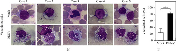
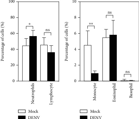
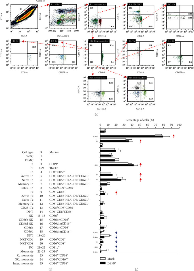
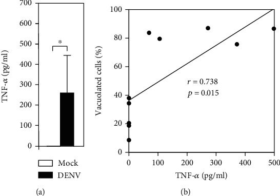
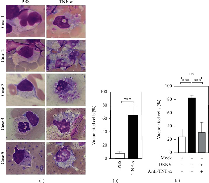
During the acute febrile phase of dengue virus (DENV) infection, viremia can cause severe systemic immune responses accompanied by hematologic disorders. This study investigated the potential induction and mechanism of the cytopathic effects of DENV on peripheral blood cells ex vivo. At one day postinfection, there was viral nonstructural protein NS1 but no further virus replication measured in the whole blood culture. Notably, DENV exposure caused significant vacuolization in monocytic phagocytes. With a minor change in the complete blood cell count, except for a minor increase in neutrophils and a significant decrease in monocytes, the immune profiling assay identified several changes, particularly a significant reduction in CD14-positive monocytes as well as CD11c-positive dendritic cells. Abnormal production of TNF-α was highly associated with the induction of vacuolization. Manipulating TNF-α expression resulted in cytopathogenic effects. These results demonstrate the potential hematological damage caused by ex vivo DENV-induced TNF-α.
Copyright © 2021 Rahmat Dani Satria et al.
Conflict of interest statement
The authors declare that there is no conflict of interest.
Figures
Similar articles
-
Differential regulation of toll-like receptor-2, toll-like receptor-4, CD16 and human leucocyte antigen-DR on peripheral blood monocytes during mild and severe dengue fever.Immunology. 2010 Jun;130(2):202-16. doi: 10.1111/j.1365-2567.2009.03224.x. Epub 2010 Jan 27. Immunology. 2010. PMID: 20113369 Free PMC article.
-
Evaluation of dengue NS1 antigen detection tests with acute sera from patients infected with dengue virus in Venezuela.Diagn Microbiol Infect Dis. 2009 Nov;65(3):247-53. doi: 10.1016/j.diagmicrobio.2009.07.022. Epub 2009 Sep 5. Diagn Microbiol Infect Dis. 2009. PMID: 19733994
-
Dengue Virus Infection with Highly Neutralizing Levels of Cross-Reactive Antibodies Causes Acute Lethal Small Intestinal Pathology without a High Level of Viremia in Mice.J Virol. 2015 Jun;89(11):5847-61. doi: 10.1128/JVI.00216-15. Epub 2015 Mar 18. J Virol. 2015. PMID: 25787279 Free PMC article.
-
Mechanisms of monocyte cell death triggered by dengue virus infection.Apoptosis. 2018 Dec;23(11-12):576-586. doi: 10.1007/s10495-018-1488-1. Apoptosis. 2018. PMID: 30267240 Review.
-
Role of Monocytes in the Pathogenesis of Dengue.Arch Immunol Ther Exp (Warsz). 2019 Feb;67(1):27-40. doi: 10.1007/s00005-018-0525-7. Epub 2018 Sep 20. Arch Immunol Ther Exp (Warsz). 2019. PMID: 30238127 Review.
Cited by
-
Elevated TNF-α Induces Thrombophagocytosis by Mononuclear Cells in ex vivo Whole-Blood Co-Culture with Dengue Virus.J Inflamm Res. 2022 Mar 5;15:1717-1728. doi: 10.2147/JIR.S356742. eCollection 2022. J Inflamm Res. 2022. PMID: 35282270 Free PMC article.
-
Cytoplasmic Vacuolization: A Fascinating Morphological Alteration From Cellular Stress to Cell Death.Cancer Sci. 2025 May;116(5):1181-1192. doi: 10.1111/cas.70013. Epub 2025 Feb 27. Cancer Sci. 2025. PMID: 40017124 Free PMC article. Review.
-
Knockout of the Tnfa Gene Decreases Influenza Virus-Induced Histological Reactions in Laboratory Mice.Int J Mol Sci. 2024 Jan 18;25(2):1156. doi: 10.3390/ijms25021156. Int J Mol Sci. 2024. PMID: 38256229 Free PMC article.
-
In vitro analysis of VEGF-mediated endothelial permeability and the potential therapeutic role of Anti-VEGF in severe dengue.Biochem Biophys Rep. 2024 Aug 22;39:101814. doi: 10.1016/j.bbrep.2024.101814. eCollection 2024 Sep. Biochem Biophys Rep. 2024. PMID: 39263317 Free PMC article.
References
-
- World Health Organization. Handbook for Clinical Management of Dengue. 2012. p. p. 114.
-
- Chaloemwong J., Tantiworawit A., Rattanathammethee T., et al. Useful clinical features and hematological parameters for the diagnosis of dengue infection in patients with acute febrile illness: a retrospective study. BMC Hematology. 2018;18(1):p. 20. doi: 10.1186/s12878-018-0116-1. - DOI - PMC - PubMed
Publication types
MeSH terms
Substances
LinkOut - more resources
Full Text Sources
Other Literature Sources
Medical
Research Materials
