

Case Reports
doi: 10.1016/j.jdcr.2021.04.006.
eCollection 2021 Jun.
Clinical features of tumor necrosis factor-α-inhibitor induced chilblain lupus: A case series
Affiliations
- PMID: 34041339
- PMCID: PMC8144103
- DOI: 10.1016/j.jdcr.2021.04.006
Item in Clipboard
Case Reports
Clinical features of tumor necrosis factor-α-inhibitor induced chilblain lupus: A case series
JAAD Case Rep.
.
No abstract available
Keywords: ANA, antinuclear antibody; ATIL, anti-TNF-α-induced lupus; CCLE, chronic cutaneous lupus erythematosus; DILE, drug-induced lupus erythematosus; SCLE, subacute cutaneous lupus erythematosus; SLE, systemic lupus erythematous; TNF inhibitor; TNF, tumor necrosis factor; anti-TNF-α-induced lupus; chilblain lupus; drug-induced lupus erythematosus; pernio; tumor necrosis factor α.
Conflict of interest statement
None declared.
Figures
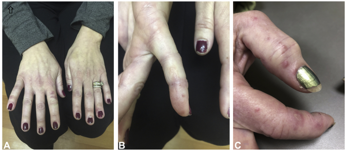
Chilblain lupus associated with TNF-α inhibitor therapy: Clinical images from cases 1 (1A, 1B) and 2 (1C) A and B, Light pink papules on the dorsal and lateral aspects of multiple fingers. C, Violaceous papules on the dorsal and lateral edges of the right first and second fingers.

Chilblain lupus associated with TNF-α inhibitor therapy: Punch biopsy specimen from Case 1 (A, B, C). Hematoxylin-eosin stains; original magnification: ×4 (A) and ×10 (B and C), demonstrating a superficial and deep perivascular and periadnexal lymphocytic infiltrate on acral skin, consistent with chilblain lupus.

Chilblain lupus associated with TNF-α inhibitor therapy: Punch biopsy specimens from cases 2 (A) and 3 (B, C). A, Alcian-blue stain, original magnification: ×10, highlighting mucin in the interstitial dermis. Hematoxylin-eosin stains, original magnification: B, ×4 and C, ×10 demonstrating a dermal perivascular and periadnexal lymphocytic infiltrate, with focal interface changes and rare necrotic keratinocytes.
Similar articles
-
Drug-induced lupus: an update on its dermatologic aspects.Lupus. 2009 Oct;18(11):935-40. doi: 10.1177/0961203309106176. Lupus. 2009. PMID: 19762393 Review.
-
Drug-induced lupus erythematosus.Arch Dermatol Res. 2009 Jan;301(1):99-105. doi: 10.1007/s00403-008-0895-5. Epub 2008 Sep 17. Arch Dermatol Res. 2009. PMID: 18797892 Review.
-
Ustekinumab-induced subacute cutaneous lupus.JAAD Case Rep. 2019 Mar 1;5(3):271-273. doi: 10.1016/j.jdcr.2019.01.015. eCollection 2019 Mar. JAAD Case Rep. 2019. PMID: 30891478 Free PMC article. No abstract available.
-
Drug-induced lupus erythematosus with emphasis on skin manifestations and the role of anti-TNFα agents.J Dtsch Dermatol Ges. 2012 Dec;10(12):889-97. doi: 10.1111/j.1610-0387.2012.08000.x. Epub 2012 Sep 3. J Dtsch Dermatol Ges. 2012. PMID: 22937775 Free PMC article. Review.
-
Comparative analysis of subacute cutaneous lupus erythematosus and chronic cutaneous lupus erythematosus: clinical and immunological study of 270 patients.Br J Dermatol. 2010 Jan;162(1):91-101. doi: 10.1111/j.1365-2133.2009.09472.x. Epub 2009 Sep 28. Br J Dermatol. 2010. PMID: 19785596
Cited by
-
Chilblain lupus induced by infliximab therapy.Rheumatol Adv Pract. 2024 Mar 5;9(2):rkaf027. doi: 10.1093/rap/rkaf027. eCollection 2025. Rheumatol Adv Pract. 2024. PMID: 40160999 Free PMC article. No abstract available.
-
A case of belatacept-induced chilblain lupus.JAAD Case Rep. 2022 Jan 22;21:112-115. doi: 10.1016/j.jdcr.2022.01.007. eCollection 2022 Mar. JAAD Case Rep. 2022. PMID: 35198715 Free PMC article. No abstract available.
References
-
- Hedrich C.M., Fiebig B., Hauck F.H. Chilblain lupus erythematosus–a review of literature. Clin Rheumatol. 2008;27(8):949–954. - PubMed
-
- Sifuentes Giraldo W.A., Ahijón Lana M., García Villanueva M.J., González García C., Vázquez Diaz M. Chilblain lupus induced by TNF-α antagonists: a case report and literature review. Clin Rheumatol. 2012;31(3):563–568. - PubMed
-
- Boada A., Bielsa I., Fernández-Figueras M.T., Ferrándiz C. Perniosis: clinical and histopathological analysis. Am J Dermatopathol. 2010;32(1):19–23. - PubMed
-
- Cribier B., Djeridi N., Peltre B., Grosshans E. A histologic and immunohistochemical study of chilblains. J Am Acad Dermatol. 2001;45(6):924–929. - PubMed
Publication types
LinkOut - more resources
Full Text Sources
Other Literature Sources