Review
. 2021 Apr;113(2):85-94.
doi: 10.32074/1591-951X-123.

Which type of cancer is detected in breast screening programs? Review of the literature with focus on the most frequent histological features

Affiliations
Review

Which type of cancer is detected in breast screening programs? Review of the literature with focus on the most frequent histological features

Angelo G Corradini et al. Pathologica. 2021 Apr.

Abstract

Breast cancer is the most frequent type of cancer affecting female patients. The introduction of breast cancer screening programs led to a substantial reduction of mortality from breast cancer. Nevertheless, doubts are being raised on the real efficacy of breast screening programs. The aim of the present paper is to review the main pathological type of cancers detected in breast cancer screening programs. Specifically, attention will be given to: in situ carcinoma, invasive carcinoma histotypes and interval cancer.

Keywords: breast cancer; in situ carcinoma; interval cancer; invasive carcinoma; screening program.


Conflict of interest statement

Conflict of interest

The Authors declare no conflict of interest.

Figures
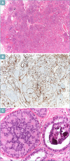
Figure 1.
Figure 1.
Low grade DCIS arising in sclerosing adenosis. (A): low power view showing closely packed glands. (B): cytokeratin 14 evidences the presence of a myoepithelial layer. (C): at higher power ducts are filled with monotonous neoplastic cells forming glandular lumina, psammomatous microcalcifications are present (arrow).
Figure 2.
Figure 2.
High grade DCIS arising in sclerosing adenosis. (A): low power view, the lesion presents a multinodular growth. (B): same lesion, stained with Cytokeratin 14 that evidences the myoepithelial layer. (C): at higher power ducts are lined by markedly atypical neoplastic cells, necrosis is present (arrow). D: cytokeratin 14 is helpful to avoid overdiagnosis of invasive carcinoma.
Figure 3.
Figure 3.
Low grade DCIS and differential diagnosis with florid epitheliosis. (A): low grade DCIS is characterized by a monotonous proliferation of neoplastic cells with bland nuclei. Neoplastic cells are polarized, with the secretroy pole oriented toward the lumen of the glandular strucure (arrow). (B): CK 14 stains the myoepithelial cells located at the periphery of the ducts, while the neoplastic cells are negative. (C): epitheliosis/usual duct hiperplasia is characterized by intraductal proliferation of cells devoid of atypia. Irregular spaces without any polarization are present. (D): in epithelosis CK 14 stains most of the intraductal cells.
Figure 4.
Figure 4.
Lobular carcinoma in situ, florid type. (A): the acinar units are filled with a solid proliferation of neoplastic cells, with uniform nuclei, necrosis is present (arrow). (B): E-Cadherin is negative in the neoplastic cells. (C): P-LCIS is characterized by enlarged terminal ductular-lobular units, filled with neoplastic cells, resembling high grade DCIS. (D): at higher power neoplastic cells are irregular. Binucleated neoplastic cells are easily detected (arrow). (E): E-Cadherin is negative, confirming the diagnosis of P-LCIS. (F): low molecular weight cytokeratins evidence small foci of invasion.
Figure 5.
Figure 5.
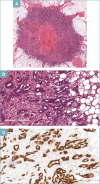
Tubular carcinoma (TC). (A): at low power view TC presents finely irregular margins. (B): at higher power, it is composed of angulated glands, lined by monotonous neoplastic cells. (C): almost all the neoplastic cells are strongly positive for estrogen receptor.
Figure 6.
Figure 6.
Invasive lobular carcinoma (ILC). (A): mammographically detected ILC associated with obliterative mastopathy. Obliterating ducts (arrow) show in situ lobular carcinoma of classical type; the surrounding tissue is infiltrated by neoplastic cells with minimal architectural distortion. (B): at higher power, neoplastic cells surround obliterating duct with in situ lobular carcinoma. (C): both in situ and invasive components are E-Cadherin negative. (D): both in situ and invasive components are strongly positive for estrogen receptor.
Figure 7.
Figure 7.
Metaplastic carcinoma. (A): at low power MC shows lobulated margins that can simulate a fibroadenoma. (B): at higher power MC is composed of markedly atypical cells, high nuclear grade in situ duct carcinoma is also present.

References

    1. Ferlay J, Shin HR, Bray F, et al. . Estimates of worldwide burden of cancer in 2008: GLO-BOCAN 2008. Int J Cancer 2010;127:2893-917. https://doi.org/10.1002/ijc.25516. 10.1002/ijc.25516 PubMed PMID: . - DOI - PubMed
    1. Bleyer A, Welch HG. Effect of three decades of screening mammography on breast-cancer in-cidence. N Engl J Med 2012;367:1998-2005. https://doi.org/10.1056/NEJMoa1206809. 10.1056/NEJMoa1206809 PubMed PMID: . - DOI - PubMed
    1. Allison K, Brogi E, Ellis IO, et al. . WHO Classification of Breast Tumours. 5th ed. Lyon: IARC Press; 2019.
    1. Park TS, Hwang ES. Current trends in the management of ductal carcinoma in situ. Oncol-ogy (Williston Park) 2016;30:823-31. PMID: . - PubMed
    1. Sidiropoulou Z, Vasconcelos AP, Couceiro C, et al. . Prevalence of silent breast cancer in autopsy specimens, as studied by the disease being held by image-guided biopsies: the pilot study and literature review. Mol Clin Oncol 2017;7:193-199. https://doi.org/10.3892/mco.2017.1299. 10.3892/mco.2017.1299 Epub 2017 Jun 21. PubMed PMID: ; PubMed Central PMCID: . - DOI - PMC - PubMed