Observational Study
. 2021 Sep 1;6(9):1078-1087.
doi: 10.1001/jamacardio.2021.2065.

Prevalence of Clinical and Subclinical Myocarditis in Competitive Athletes With Recent SARS-CoV-2 Infection: Results From the Big Ten COVID-19 Cardiac Registry

Collaborators, Affiliations
Observational Study

Prevalence of Clinical and Subclinical Myocarditis in Competitive Athletes With Recent SARS-CoV-2 Infection: Results From the Big Ten COVID-19 Cardiac Registry

Curt J Daniels et al. JAMA Cardiol. .

Abstract

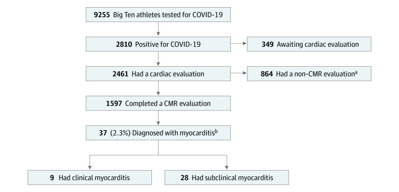
Importance: Myocarditis is a leading cause of sudden death in competitive athletes. Myocardial inflammation is known to occur with SARS-CoV-2. Different screening approaches for detection of myocarditis have been reported. The Big Ten Conference requires comprehensive cardiac testing including cardiac magnetic resonance (CMR) imaging for all athletes with COVID-19, allowing comparison of screening approaches.

Objective: To determine the prevalence of myocarditis in athletes with COVID-19 and compare screening strategies for safe return to play.

Design, setting, and participants: Big Ten COVID-19 Cardiac Registry principal investigators were surveyed for aggregate observational data from March 1, 2020, through December 15, 2020, on athletes with COVID-19. For athletes with myocarditis, presence of cardiac symptoms and details of cardiac testing were recorded. Myocarditis was categorized as clinical or subclinical based on the presence of cardiac symptoms and CMR findings. Subclinical myocarditis classified as probable or possible myocarditis based on other testing abnormalities. Myocarditis prevalence across universities was determined. The utility of different screening strategies was evaluated.

Exposures: SARS-CoV-2 by polymerase chain reaction testing.

Main outcome and measure: Myocarditis via cardiovascular diagnostic testing.

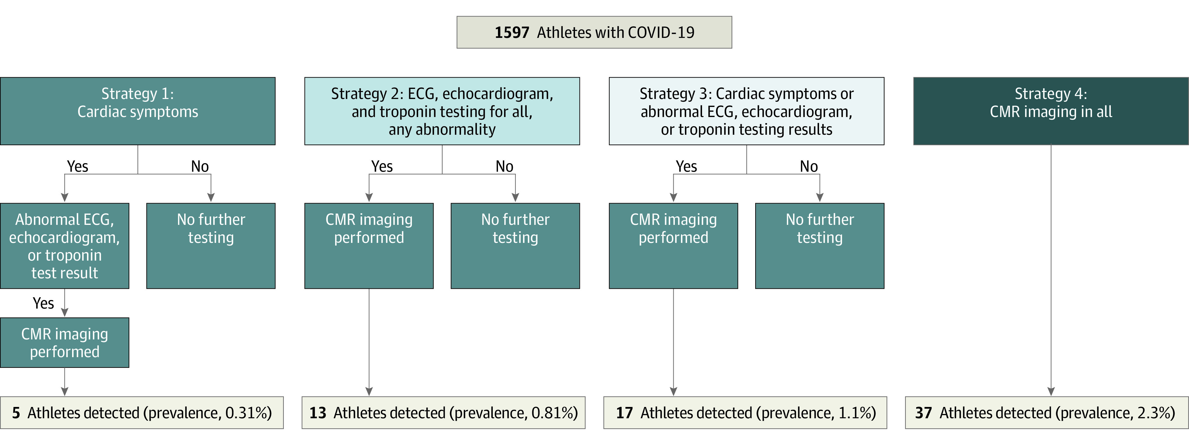
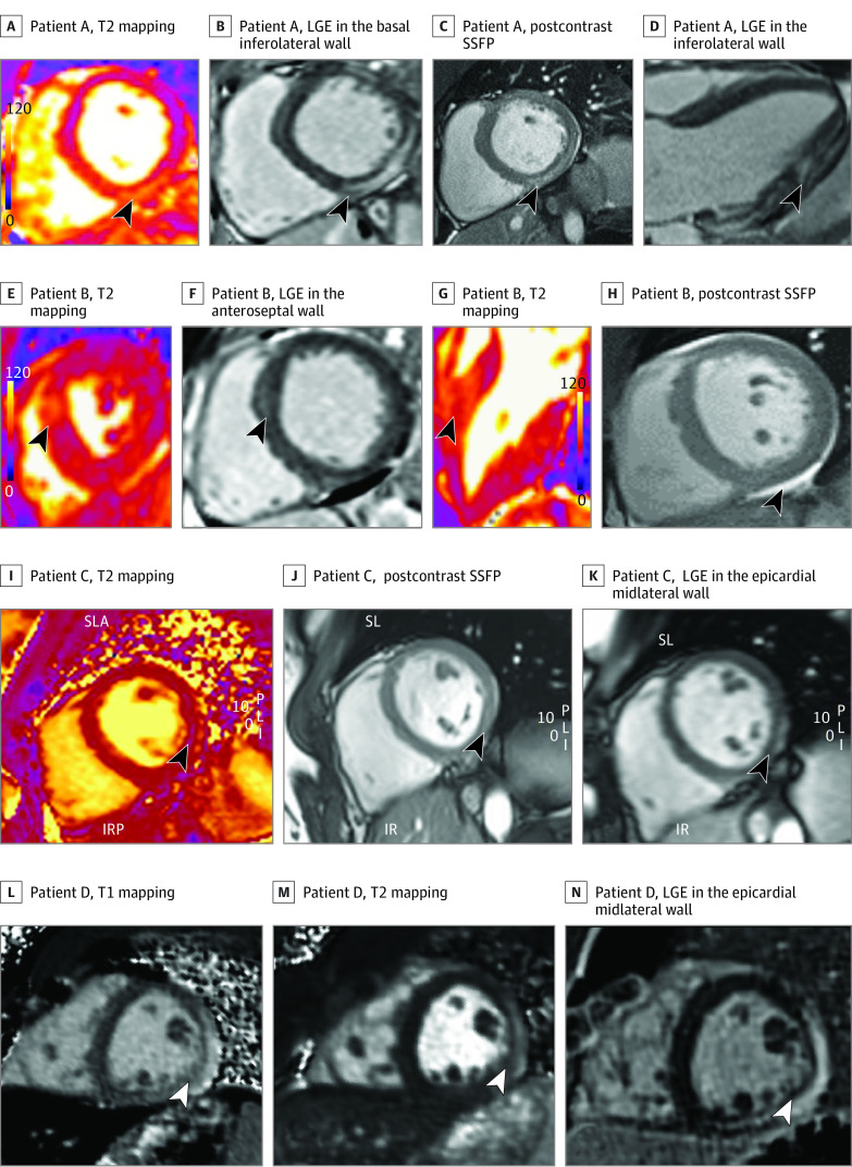
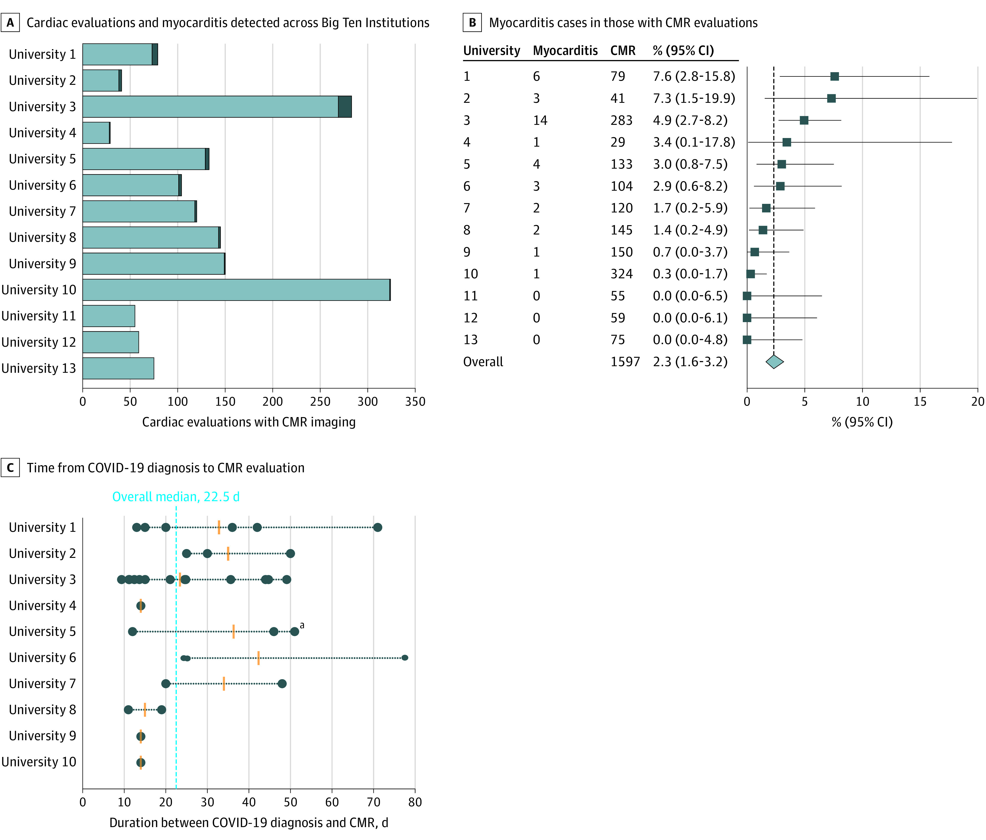
Results: Representing 13 universities, cardiovascular testing was performed in 1597 athletes (964 men [60.4%]). Thirty-seven (including 27 men) were diagnosed with COVID-19 myocarditis (overall 2.3%; range per program, 0%-7.6%); 9 had clinical myocarditis and 28 had subclinical myocarditis. If cardiac testing was based on cardiac symptoms alone, only 5 athletes would have been detected (detected prevalence, 0.31%). Cardiac magnetic resonance imaging for all athletes yielded a 7.4-fold increase in detection of myocarditis (clinical and subclinical). Follow-up CMR imaging performed in 27 (73.0%) demonstrated resolution of T2 elevation in all (100%) and late gadolinium enhancement in 11 (40.7%).

Conclusions and relevance: In this cohort study of 1597 US competitive athletes with CMR screening after COVID-19 infection, 37 athletes (2.3%) were diagnosed with clinical and subclinical myocarditis. Variability was observed in prevalence across universities, and testing protocols were closely tied to the detection of myocarditis. Variable ascertainment and unknown implications of CMR findings underscore the need for standardized timing and interpretation of cardiac testing. These unique CMR imaging data provide a more complete understanding of the prevalence of clinical and subclinical myocarditis in college athletes after COVID-19 infection. The role of CMR in routine screening for athletes safe return to play should be explored further.


Conflict of interest statement

Conflict of Interest Disclosures: Dr Daniels reported a donation from a family fund to support the research team and regulatory work at Ohio State University. Dr Rajpal reported grants from the Jay and Jeanie Schottenstein Foundation during the conduct of the study. Dr Albers reported that their institution, the University of Nebraska, applied for $5000 from the Big Ten Conference Cardiac Registry to cover expenses incurred in the collection and submission of data; if received by the University of Nebraska, Dr Albers will not receive any of these funds. Dr Day reported support for regulatory costs from Indiana University to their institution. Dr Somers reported grants from the National Institutes of Health during the conduct of the study. No other disclosures were reported.

Figures

Figure 1.
Figure 1.. Cohort of Big Ten Athletes
aAthletes were excluded from analysis for not completing cardiac magnetic resonance (CMR) imaging as part of cardiac evaluation and described in more detail in eAppendix 2 in Supplement 1. bAthletes diagnosed with myocarditis were categorized as clinical or subclinical based on presence or absence of cardiac symptoms.
Figure 2.
Figure 2.. Detection and Estimated Prevalence of Myocarditis Based on Diagnostic Strategy
From 37 athletes with clinical and subclinical myocarditis, the number that would have been detected and percentage prevalence found based on strategy performed and guided by either (1) cardiac symptoms alone; (2) electrocardiogram (ECG), echocardiogram, and troponin for all; (3) cardiac symptoms, ECG, echocardiogram, or troponin; or (4) cardiovascular magnetic resonance (CMR) imaging for all strategy.
Figure 3.
Figure 3.. Cardiac Magnetic Resonance Imaging in Athletes With Clinical and Subclinical Myocarditis
A-D, Athlete A with subclinical possible myocarditis was asymptomatic with normal electrocardiogram (ECG), echocardiogram, and high-sensitivity troponin findings. A, T2 mapping showing elevated T2 in basal-mid inferolateral wall in short axis view. B, late gadolinium enhancement (LGE) in the basal inferolateral wall in short axis view. C, Postcontrast steady state-free precession (SSFP) images showing contrast uptake in the basal-mid inferolateral wall in short axis view. D, LGE in the inferolateral wall in 3-chamber view. E-H, Athlete B with subclinical probable myocarditis was asymptomatic with normal ECG, normal echocardiogram, and elevated high-sensitivity troponin findings. E, T2 mapping showing elevated T2 in the anteroseptal wall in short axis view. F, LGE in the anteroseptal wall in 3-chamber view. G, T2 mapping showing elevated T2 in the anteroseptal wall in 3-chamber view. F, Postcontrast SSFP image showing pericardial effusion in short axis view. I-K, Athlete C with clinical myocarditis and chest pain, dyspnea, abnormal ECG, normal echocardiogram, and normal troponin findings. I, T2 mapping showing elevated T2 in the lateral wall short axis view. J, Postcontrast SSFP images showing contrast uptake in midlateral wall in short axis view. K, LGE in the epicardial midlateral wall in short axis view. L-N, Athlete D with clinical myocarditis, chest pain, abnormal ECG, echocardiogram, and troponin findings. L, T1 mapping showing elevated native T1 in midlateral wall in short axis view. M, T2 mapping showing elevated T2 in the midlateral wall in short axis view. N, LGE in the epicardial midlateral wall in short axis view. IR indicates inferior right view; IRP, inferior, right, posterior view; PLI, posterior, left, inferior view; SL, superior left view; SLA, superior, left, anterior view.
Figure 4.
Figure 4.. Cardiac Evaluations Performed in Big Ten Athletes
Cardiovascular magnetic resonance (CMR) imaging, athletes diagnosed with myocarditis, and CMR timing after COVID-19 diagnosis in Big Ten athletes with recent SARS-CoV-2 infection. A, The reported number of normal (light blue) and athletes diagnosed with myocarditis (dark blue) observed from complete cardiac evaluations including CMR imaging completed in Big Ten athletes with recent SARS-CoV-2 infection. B, The reported number of athlethes diagnosed with myocarditis, complete cardiac evaluations including CMR imaging, and the percent myocarditis with the 95% CI (calculated using the Clopper-Pearson exact method) for each participating university and the overall prevalence (crude estimate, calculated as the quotient of all athletes with myocarditis and CMR imaging performed across all universities). C, Duration between COVID-19 diagnosis and CMR imaging for athletes who were diagnosed with myocarditis (n = 36). Data are displayed for participating institution where cases of myocarditis were observed (n = 10). aDuration data unknown for 1 athlete diagnosed with myocarditis. Filled circles represent individual case duration, and orange horizontal lines represent the institution median duration between COVID-19 diagnosis and CMR imaging.

Comment in

  • doi: 10.1001/jamacardio.2021.2079

Similar articles

Cited by

References

    1. Singh R, Kang A, Luo X, et al. . COVID-19: current knowledge in clinical features, immunological responses, and vaccine development. FASEB J. 2021;35(3):e21409. doi:10.1096/fj.202002662R - DOI - PMC - PubMed
    1. Corrado D, Zorzi A. Sudden death in athletes. Int J Cardiol. 2017;237:67-70. doi:10.1016/j.ijcard.2017.03.034 - DOI - PubMed
    1. Finocchiaro G, Papadakis M, Robertus JL, et al. . Etiology of sudden death in sports: insights from a United Kingdom regional registry. J Am Coll Cardiol. 2016;67(18):2108-2115. doi:10.1016/j.jacc.2016.02.062 - DOI - PubMed
    1. Peterson DF, Kucera K, Thomas LC, et al. . Aetiology and incidence of sudden cardiac arrest and death in young competitive athletes in the USA: a 4-year prospective study. Br J Sports Med. Published online November 12, 2020. doi:10.1136/bjsports-2020-102666 - DOI - PMC - PubMed
    1. Harmon KG, Asif IM, Maleszewski JJ, et al. . Incidence, cause, and comparative frequency of sudden cardiac death in national collegiate athletic association athletes: a decade in review. Circulation. 2015;132(1):10-19. doi:10.1161/CIRCULATIONAHA.115.015431 - DOI - PMC - PubMed

Publication types