. 2021 Oct;32(5):332-349.
doi: 10.1007/s00335-021-09875-3. Epub 2021 May 27.

A comprehensive phenotypic characterization of a whole-body Wdr45 knock-out mouse

Affiliations

A comprehensive phenotypic characterization of a whole-body Wdr45 knock-out mouse

Caroline A Biagosch et al. Mamm Genome. 2021 Oct.

Abstract

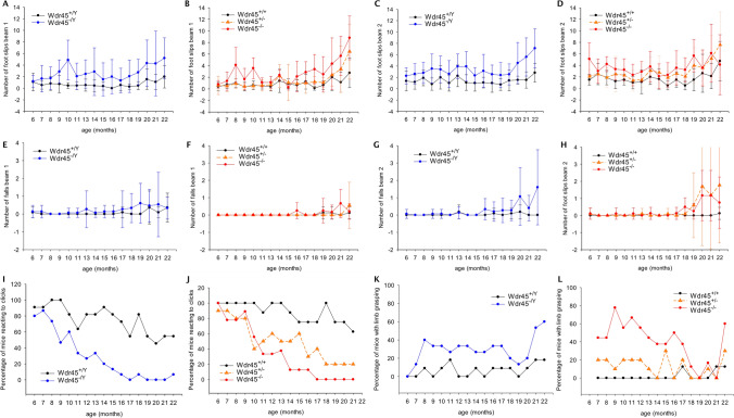
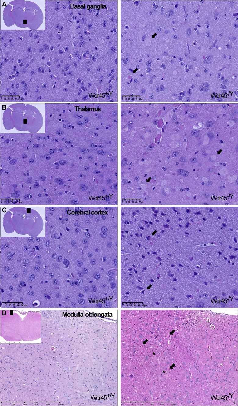
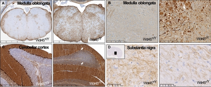
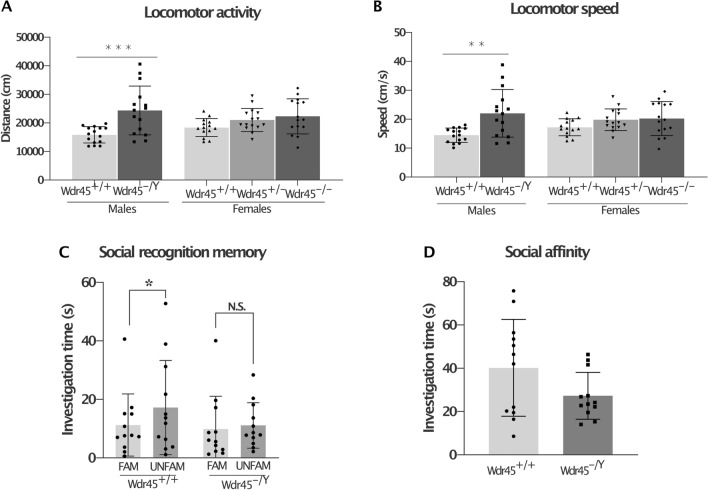
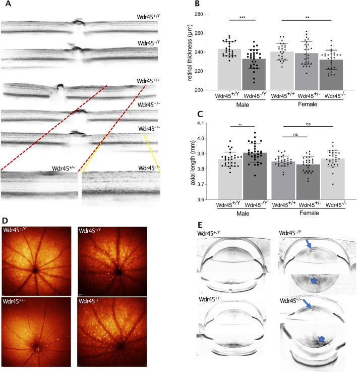
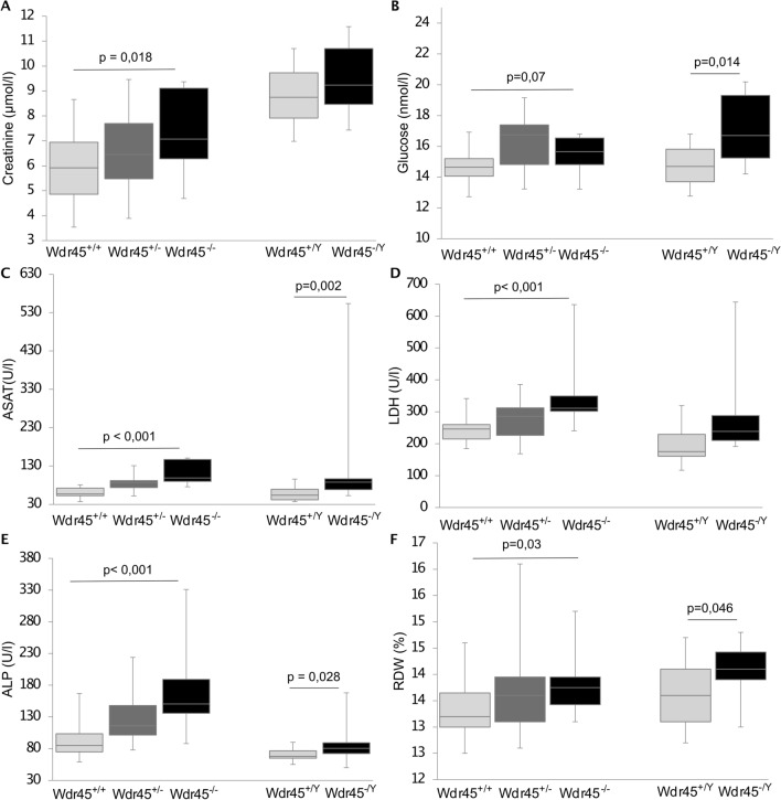
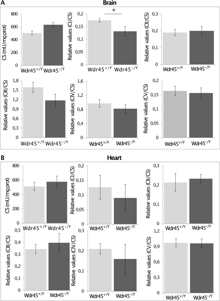
Pathogenic variants in the WDR45 (OMIM: 300,526) gene on chromosome Xp11 are the genetic cause of a rare neurological disorder characterized by increased iron deposition in the basal ganglia. As WDR45 encodes a beta-propeller scaffold protein with a putative role in autophagy, the disease has been named Beta-Propeller Protein-Associated Neurodegeneration (BPAN). BPAN represents one of the four most common forms of Neurodegeneration with Brain Iron Accumulation (NBIA). In the current study, we generated and characterized a whole-body Wdr45 knock-out (KO) mouse model. The model, developed using TALENs, presents a 20-bp deletion in exon 2 of Wdr45. Homozygous females and hemizygous males are viable, proving that systemic depletion of Wdr45 does not impair viability and male fertility in mice. The in-depth phenotypic characterization of the mouse model revealed neuropathology signs at four months of age, neurodegeneration progressing with ageing, hearing and visual impairment, specific haematological alterations, but no brain iron accumulation. Biochemically, Wdr45 KO mice presented with decreased complex I (CI) activity in the brain, suggesting that mitochondrial dysfunction accompanies Wdr45 deficiency. Overall, the systemic Wdr45 KO described here complements the two mouse models previously reported in the literature (PMIDs: 26,000,824, 31,204,559) and represents an additional robust model to investigate the pathophysiology of BPAN and to test therapeutic strategies for the disease.


Conflict of interest statement

The authors state no conflict of interest.

Figures

Fig. 1
Fig. 1
Neurologic deterioration in Wdr45 KO mice. ad Males Wdr45−/Y (A, C), as well as females Wdr45−/− (b, d) of the progression cohort, made more slips compared to controls on both beams tested (linear mixed-effects model; genotype effect p < 0.001 each). eh The number of falls from the different beams was not altered on beam 1 (e, f) but mildly increased for beam 2 (g, h; linear mixed-effects model; genotype effects Wdr45−/Y p < 0.05; Wdr45−/+ p < 0.05; Wdr45−/− n.s.). i, j) Decreasing numbers of both sexes’ mutant mice reacted to a clickbox (linear model; genotype p < 0.001 each). k, l) More mutants showed limb grasping compared to controls (linear model; genotype effects p < 0.001 for Wdr45−/Y and Wdr45−/−, p < 0.05 for Wdr45−/+). The number of mice used for the tests was as follows: Wdr45−/+ n = 10, Wdr45−/− n = 9, Wdr45+/+ n = 8, Wdr45−/Y n = 15, Wdr45+/Y n = 11
Fig. 2
Fig. 2
H&E staining in representative tissue sections from 18-month-old Wdr45+/Y and Wdr45−/Y mice. ad Black squares in the insets indicate the relevant brain area zoomed in the figure. Arrows indicate eosinophil spheroids, while asterisks indicate swollen structures potentially showing degenerated neurons in basal ganglia, thalamus, cerebral cortex, and medulla oblongata of Wdr45 KO mice. Scale bar represents 50 µm for figures A-C; 500 µm for D
Fig. 3
Fig. 3
Immunostaining in representative tissue sections from 18-month-old Wdr45 +/Y and Wdr45−/Y mice. a GFAP staining and b Ubiquitin staining in medulla oblongata; c Calbindin staining of the cerebellar cortex; d Dopamine staining of substantia nigra. Scale bar represents 50 µm for figures A- B and 500 µm for figure D
Fig. 4
Fig. 4
Locomotor activity and social discrimination in Wdr45 KO mice. There was increased total distance travelled a and locomotor speed b by the male Wdr45−/Y mice in the open field at 11 months of age. A similar pattern was visible in especially the female Wdr45−/− mice. **p < 0.01, ***p < 0.001, 1-way ANOVA with post hoc Tukey’s test. Social discrimination memory was impaired in the male Wdr45−/Y mice at 12.5 months of age (c). During the test phase, the Wdr45−/Y mice spent equivalent times investigating familiar (“FAM”) vs unfamiliar (“UNFAM”) stimulus animals, while the control mice spent significantly more time investigating UNFAM mice. A pattern of decreased social affinity was also evident (d). Bars indicate standard deviation; *paired t test: p < 0.05. The number of mice used for the tests was as follows: open field: male Wdr45+/+ n = 15, Wdr45−/Y n = 15, female Wdr45+/+ n = 14, Wdr45± n = 15, Wdr45−/− n = 15; social discrimination: male Wdr45+/+ n = 12, Wdr45−/Y n = 12
Fig. 5
Fig. 5
Ophthalmic examination in Wdr45 KO mice. a SD-OCT imaging of the retinal layers indicating granular-like alterations in the retinal layers of the female Wdr45−/− and male Wdr45−/Y mice from the progression cohort. Magnification of the granular-like alterations in the insets. b SD-OCT evaluation of the total retinal thickness. c Biometric measurement of the eye-axial lengths. The bar charts show pooled data of both eyes for each animal. The genotype effect was evaluated with the Wilcoxon rank-sum test (homozygous females/hemizygous males vs wild-type). d BAF funduscopy of the inner retinal layers in female Wdr45−/− and male Wdr45−/y mice from the F8–F9 generations. Accumulation of hyperfluorescent profiles is evident. e SD-OCT images of the eye lens in female Wdr45−/− and male Wdr45−/y mice from the F8–F9 generations showing an accumulation of protein aggregates in the lens anterior cortex (arrow). Additional alterations are seen as darker spots in the posterior lens cortex (arrowheads). The number of mice used for the tests was as follows: Wdr45+/+ n = 12, Wdr45−/− n = 13 Wdr45 +/Y n = 13, and Wdr45 −/Y n = 15 from the F4 cohort; Wdr45+/+ n = 6, Wdr45−/− n = 2, Wdr45 + −/Y n = 3, and Wdr45 −/Y n = 5 from the F8–F9 cohort
Fig. 6
Fig. 6
Clinical chemistry and haematology investigation. a Creatinine, b glucose, c aspartate aminotransferase (ASAT), d lactate dehydrogenase (LDH), e alkaline phosphatase (ALP), and f red blood cells distributed widths (RDW) were measured in 14-month-old mice. Tests for genotype effects were made using the Wilcoxon rank-sum test (homozygous females/hemizygous males vs wild-type). The number of mice used for the tests was as follows: Wdr45−/+ n = 15, Wdr45−/− n = 14, Wdr45+/+ n = 15, Wdr45−/Y n = 14, Wdr45+/Y n = 15
Fig. 7
Fig. 7
The respiratory chain complexes I–V activity and the citrate synthase (CS) measured in a brain and b heart of mice from the progression cohort. Data on the activity of the complexes I–V are normalized to the CS. Data are represented as mean ± SEM. The number of mice used for the brain’s respiratory chain activities measurements was n = 6, while for the measures in the heart, mice were n = 3. Mann–Whitney test, *p < 0.05

References

    1. Abidi A, Mignon-Ravix C, Cacciagli P, Girard N, Milh M, Villard L. Early-onset epileptic encephalopathy as the initial clinical presentation of WDR45 deletion in a male patient. Eur J Hum Genet. 2016;24(4):615–618. doi: 10.1038/ejhg.2015.159. - DOI - PMC - PubMed
    1. André V, Gau C, Scheideler A, Aguilar-Pimentel JA, Amarie OV, Becker L, Garrett L, Hans W, Hölter SM, Janik D, Moreth K, Neff F, Östereicher M, Racz I, Rathkolb B, Rozman J, Bekeredjian R, Graw J, Klingenspor M, Klopstock T, Ollert M, Schmidt-Weber C, Wolf E, Wurst W, Gailus-Durner V, Brielmeier M, Fuchs H, Hrabé de Angelis M. Laboratory mouse housing conditions can be improved using common environmental enrichment without compromising data. PLoS Biol. 2018;16(4):e2005019. doi: 10.1371/journal.pbio.2005019. - DOI - PMC - PubMed
    1. Behrends C, Sowa ME, Gygi SP, Harper JW. Network organization of the human autophagy system. Nature. 2010;466(7302):68–76. doi: 10.1038/nature09204. - DOI - PMC - PubMed
    1. Belohlavkova A, Sterbova K, Betzler C, Burkhard S, Panzer A, Wolff M, Lassuthova P, Vlckova M, Kyncl M, Benova B, Jahodova A, Kudr M, Goerg M, Dusek P, Seeman P, Kluger G, Krsek P. Clinical features and blood iron metabolism markers in children with beta-propeller protein associated neurodegeneration. Eur J Paediatr Neurol. 2020;28:81–88. doi: 10.1016/j.ejpn.2020.07.010. - DOI - PubMed
    1. Berger A, Mayr JA, Meierhofer D, Fötschl U, Bittner R, Budka H, Grethen C, Huemer M, Kofler B, Sperl W. Severe depletion of mitochondrial DNA in spinal muscular atrophy. Acta Neuropathol (berl) 2003;105(3):245–251. doi: 10.1007/s00401-002-0638-1. - DOI - PubMed

Publication types