Review
. 2021 Sep;9(5):1369-1382.
doi: 10.1111/andr.13057. Epub 2021 Jun 11.

Use of contrast enhanced ultrasound in testicular diseases: A comprehensive review

Affiliations
Review

Use of contrast enhanced ultrasound in testicular diseases: A comprehensive review

Marta Tenuta et al. Andrology. 2021 Sep.

Abstract

Background: Contrast-enhanced ultrasound (CEUS) is a sonographic technique that increases the diagnostic accuracy of ultrasound and color Doppler ultrasound (CDUS) when studying testicular abnormalities. However, its role in clinical practice is still debatable because there are no accepted standards regarding how and when this technique should be used for patients with testicular disease.

Objectives: To perform a nonsystematic review of the current literature to highlight the strength and flaws of performing CEUS and to provide a critical overview of current research evidence on this topic.

Materials and methods: A thorough search of published peer-reviewed studies in PubMed was performed using proper keywords.

Results: Strong enhancement of neoplastic lesions (both benign and malignant) during CEUS aids in differential diagnosis with non-neoplastic lesions, which usually appears either nonenhanced or enhanced in a manner similar to that of the surrounding parenchyma. CEUS enhancement has a high predictive value in the identification of neoplastic lesions, whereas a similar or complete absence of enhancement may be interpreted as strong evidence of benignity, although there are exceptions. Literature on quantitative analysis is still scarce, though promising, particularly in distinguishing benign from malignant neoplasms. Furthermore, CEUS may be useful in many emergency situations, such as acute scrotum, blunt scrotal trauma, and focal infarction of the testis. Finally, CEUS can help increase the probability of sperm recovery in azoospermic males.

Discussion and conclusion: CEUS is a safe, easy-to-perform, and cost-effective diagnostic tool that can provide a more accurate diagnosis in testicular lesions and acute scrotal disease. However, further studies with larger cohorts are required to refine the differential diagnosis between benign and malignant neoplasms. Finally, these preliminary results can instigate the development of innovative research on pre-testicular sperm extraction to increase the chances of sperm recovery.

Keywords: CEUS; acute scrotum; infertility; testicular tumor; testis.


Conflict of interest statement

The authors declare no conflict of interest regarding the publication of this article.

Figures

FIGURE 1
FIGURE 1
Qualitative analysis of CEUS. B‐mode US demonstrates a small hypoechoic lesion, with hyperechoic and well‐defined margins, resulted a Leydig cell tumor at histology. Color Doppler US demonstrates vascularity within the lesion. With contrast‐enhanced US, the lesion demonstrates marked hyperenhancement, a characteristic that has the potential to differentiate neoplastic from nonneoplastic lesions
FIGURE 2
FIGURE 2
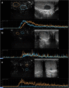
Time intensity curves (TIC). They are bell‐shaped curves that describe an initial uptake phase of the contrast medium (wash‐in) up to the maximum peak of intensity, and a subsequent release phase (washout). Lesion and parenchyma kinetics can be measured, and the resulting curves can be compared. In the figure the blue curve describes the wash‐in and wash‐out phases of a Leydig cell tumor, the orange curve describes the phases of the adjacent, normal parenchyma
FIGURE 3
FIGURE 3
Graphic representation of the time/intensity curve and the calculated perfusion parameters—wash‐in time (W‐in): the time when testis enhancement first occurs, measured in seconds; time to peak (TTP): the time needed to reach the peak intensity (PI), measured in seconds; mean transit time (MTT) or rise time (RT): the time difference between the time needed to reach the PI and the time since the beginning of ROI enhancement, measured in seconds; PI or peak enhancement (PE): the maximum ROI enhancement, measured in decibel (dB) or acoustic units (au); washout time (T‐out): the time difference between the 50% PI values in the washout and peak intensity value, measured in seconds (several studies also consider T‐out as the time needed for the descending slope to reach a contrast signal intensity of zero); area under the curve (AUC): intensities of the entire enhancement period, measured in dB or au. Several studies also differentiate wash‐in AUC (before PI) and washout AUC (after PI); slope in or β: the coefficient of the wash‐in slope, it reflects the mean blood flow velocity in the region of interest, measured in dB or au
FIGURE 4
FIGURE 4
CEUS in the differential diagnosis between non‐neoplastic and neoplastic intratesticular lesions. (a) A hyperenhanced lesion compared to the adjacent parenchyma, turned out to be a seminoma at histology. (b) A hysoenhanced lesion compared to the parenchyma resulted a Leydig cell hyperplasia at histology. (c) A hypoenhanced lesion, resulted focal fibrosis at definitive histology
FIGURE 5
FIGURE 5
Epidermoid cyst. B‐mode US demonstrates a well‐circumscribed, solid, mixed‐reflectivity lesion with high‐reflectivity “onion‐skin” peripheral rims. Contrast‐enhanced US demonstrates a clear lack of enhancement within the lesion
FIGURE 6
FIGURE 6
Quantitative analysis comparison of a seminoma and a Leydig cell tumor. A rapid wash‐in and wash‐out are distinctive characteristics of seminomas, as demonstrated by the blue curve in panel (a) that shows a wash‐in that begins approximately at 18 s and a wash‐out starting at 30–35 s, whereas a rapid wash‐in (20 s) and a delayed wash‐out (starting at 38–40 s) are appropriate signs of a Leydig cell tumor (panel b, blue curve). The orange curves belong to the adjacent normal parenchyma
FIGURE 7
FIGURE 7
Testicular torsion. CEUS showed complete lack of enhancement of testis and spermatic cord in a patient with chronic (missed) torsion. Peri‐testicular tissues displayed increased vascularity on CEUS
FIGURE 8
FIGURE 8
Intratesticular hematoma. CEUS appearances of intratesticular hematoma after blunt trauma. Dual‐display image showing contrast‐specific (left) and low MI B‐mode image (right). B‐mode image shows an intratesticular bilobated hypoechoic, avascular lesion. CEUS confirmed the absence of internal vascularity. Note the peri‐lesional hyperemia and the presence of internal echoes, representing artifact from echogenic content
FIGURE 9
FIGURE 9
Focal ischemia. CEUS appearances of focal ischemia, confirmed at definitive histology. Dual‐display image showing contrast‐specific (left) and low MI B‐mode image (right). B‐mode image shows an intratesticular, well‐defined markedly hypoechoic, avascular lesion. CEUS confirmed the complete absence of internal vascularity and the patient, monorchid, underwent tissue sparing surgery

References

    1. Lotti F, Frizza F, Balercia G, et al. The European Academy of Andrology (EAA) ultrasound study on healthy, fertile men: scrotal ultrasound reference ranges and associations with clinical, seminal, and biochemical characteristics. Andrology. 2021;9(2):559–576. - PubMed
    1. Pozza C, Kanakis G, Carlomagno F, et al. Testicular ultrasound score: a new proposal for a scoring system to predict testicular function. Andrology. 2020;8(5):1051–1063. - PMC - PubMed
    1. Dieckmann KP, Frey U, Lock G. Contemporary diagnostic work‐up of testicular germ cell tumours. Nat Rev Urol. 2013;10(12):703–712. - PubMed
    1. Pozza C, Gianfrilli D, Fattorini G, et al. Diagnostic value of qualitative and strain ratio elastography in the differential diagnosis of non‐palpable testicular lesions. Andrology. 2016;4(6):1193–1203. - PMC - PubMed
    1. Manganaro L, Saldari M, Pozza C, et al. Dynamic contrast‐enhanced and diffusion‐weighted MR imaging in the characterisation of small, non‐palpable solid testicular tumours. Eur Radiol. 2018;28(2):554–564. - PubMed