Review
. 2021 May;31(3):e12954.
doi: 10.1111/bpa.12954.

MRI and muscle imaging for idiopathic inflammatory myopathies

Affiliations
Review

MRI and muscle imaging for idiopathic inflammatory myopathies

Samuel Malartre et al. Brain Pathol. 2021 May.

Abstract

Although idiopathic inflammatory myopathies (IIM) are a heterogeneous group of diseases nearly all patients display muscle inflammation. Originally, muscle biopsy was considered as the gold standard for IIM diagnosis. The development of muscle imaging led to revisiting not only the IIM diagnosis strategy but also the patients' follow-up. Different techniques have been tested or are in development for IIM including positron emission tomography, ultrasound imaging, ultrasound shear wave elastography, though magnetic resonance imaging (MRI) remains the most widely used technique in routine. Whereas guidelines on muscle imaging in myositis are lacking here we reviewed the relevance of muscle imaging for both diagnosis and myositis patients' follow-up. We propose recommendations about when and how to perform MRI on myositis patients, and we describe new techniques that are under development.

Keywords: MRI; idiopathic inflammatory myopathies; ultra sound imaging.


Figures

FIGURE 1
FIGURE 1
High muscle T2 signals in myositis: (A) Muscle MRI of an overlap myositis (scleroderma) showing mild muscular inflammation. Normal muscles appear with a very low signal (close to the subcutaneous tissue after fat signal suppression). Inflammatory muscles exhibit high signal (slight) with blurred borders, clustered along the aponeuroses and muscular septa (full arrows). The dashed arrows show an area with a more intense hypersignal. (B) Myositis with moderate to severe muscle inflammation. MRI of a dermatomyositis patient showing hyperintense areas affecting mainly the four heads of the quadriceps muscle. (C) ASyS patient with severe muscle inflammation, displaying a marked T2 hyperintensity affecting all three compartments of the thighs on both sides. (D) MRI of an IMNM patient showing that hypersignal is also present when muscle fiber necrosis occurs in absence of significant inflammatory cell infiltratation. All pictures show thigh muscles MRI images (axial plane, T2 STIR w. seq). AM, Adductor magnus; LB, long biceps femoris; Q, quadriceps muscle; RF, rectus femoris; ST, semi‐tendinosus hamstring muscle; VI, vastus intermedius; VL, vastus lateralis; VM, vastus intermedialis
FIGURE 2
FIGURE 2
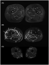
High fascia T2 signals in myositis: (A) patient with an eosinophilic fasciitis displays a diffuse hypersignal of the deep fascia and intermuscular septa (small arrows). The pelvic muscles (i.e., gluteus muscles) are also affected. (B) MRI of a patient with a graft versus host disease involving the fascia (big arrows) (diffuse hyperintensity and thickening of the deep fascia and intermuscular septa) and the muscles (especially both the adductor magnus muscles and the right quadriceps muscle). (C) ASyS patient dysplaying a fasciitis with hyperintense, thickened fascia and intermuscular septa on both sides with symmetrical distribution (full arrows). In addition, presence of a mild myositis attested by a blurred, slight T2 hyperintensity in the quadriceps muscles (dashed arrows). All pictures show thigh muscles MRI images (axial plane, T2 STIR w. seq). RF, rectus femori; VL, vastus lateralis
FIGURE 3
FIGURE 3
High muscle T2 signals in non‐myositis patients: (A) Neurogenic muscle edema was observed on an MRI performed 4 months after an iatrogenic lesion of the femoral nerve (inguinal hernia surgery). Intense signal and atrophy of the left quadriceps corresponding to the femoral nerve territory are present. Atrophy can be easily identified as compared to the contralateral thigh. Slackness of intramuscular septa (arrow) can also give a clue. (B) MRI 24 h after intense exercise showing patchy areas with increased T2 intensity affecting the quadriceps muscle (arrows), predominantly its medial head, with normal muscle biopsy and spontaneous remission of muscle signs and CK elevation. All pictures show thigh muscles MRI images (axial plane, T2 STIR w. seq). RF, rectus femoris; VI, vastus intermedius; VL, vastus lateralis; VM, vastus medialis
FIGURE 4
FIGURE 4
MRI muscle damages in myositis patients. (A) Upper limbs muscle damages in inclusion body myositis attested by a mild fatty replacement of the muscles of the forearm, involving predominantly the deep flexor digitorum (full arrows), and to a lesser extent the extensors (dashed arrows). (B) Lower limbs muscle damages in inclusion body myositis attested by a distal involvement encompassing muscle atrophy (loss of volume with the widening of the fat tissue between muscles, dashed arrows) and fatty replacement (muscular T1 hypersignal) occurring mainly in the quadriceps femori muscle (full arrows). All pictures show axial plane, T1 w. seq of the arm A or the thigh B

References

    1. Mariampillai K, Granger B, Amelin D, Guiguet M, Hachulla E, Maurier F, et al. Development of a new classification system for idiopathic inflammatory myopathies based on clinical manifestations and myositis‐specific autoantibodies. JAMA Neurol. 2018;75(12):1528–37. - PMC - PubMed
    1. Hoogendijk JE, Amato AA, Lecky BR, Choy EH, Lundberg IE, Rose MR, et al. 119th ENMC international workshop: trial design in adult idiopathic inflammatory myopathies, with the exception of inclusion body myositis, 10–12 October 2003, Naarden, The Netherlands. Neuromuscul Disord. 2004;14:337–45. - PubMed
    1. Lundberg IE, Tjärnlund A, Bottai M, Werth VP, Pilkington C, de Visser M, et al. 2017 European league against rheumatism/American College of Rheumatology classification criteria for adult and juvenile idiopathic inflammatory myopathies and their major subgroups. Arthritis Rheumatol. 2017;69:2271–82. - PMC - PubMed
    1. Aggarwal R, Rider LG, Ruperto N, Bayat N, Erman B, Feldman BM, et al. 2016 American College of Rheumatology/European league against rheumatism criteria for minimal, moderate, and major clinical response in adult dermatomyositis and polymyositis: an International Myositis Assessment and Clinical Studies Group/Paediatric Rheumatology International Trials Organisation Collaborative Initiative. Ann Rheum Dis. 2017;76:792–801. - PMC - PubMed
    1. Allenbach Y, Mammen AL, Benveniste O, Stenzel W, Immune‐Mediated Necrotizing Myopathies Working Group . 224th ENMC International Workshop: clinico‐sero‐pathological classification of immune‐mediated necrotizing myopathies Zandvoort, The Netherlands, 14‐16 October 2016. Neuromuscul Disord. 2018;28(1):87–99. - PubMed