

Cryopreservation Protocols for Genetically Engineered Mice
- PMID: 34043268
- PMCID: PMC8211118
- DOI: 10.1002/cpz1.138
Cryopreservation Protocols for Genetically Engineered Mice
Erratum in
-
Group Correction Statement (Human or Animal Subject Note).Curr Protoc. 2022 Aug;2(8):e554. doi: 10.1002/cpz1.554. Curr Protoc. 2022. PMID: 36005901 No abstract available.
Abstract
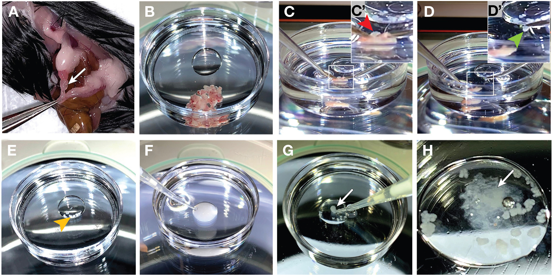
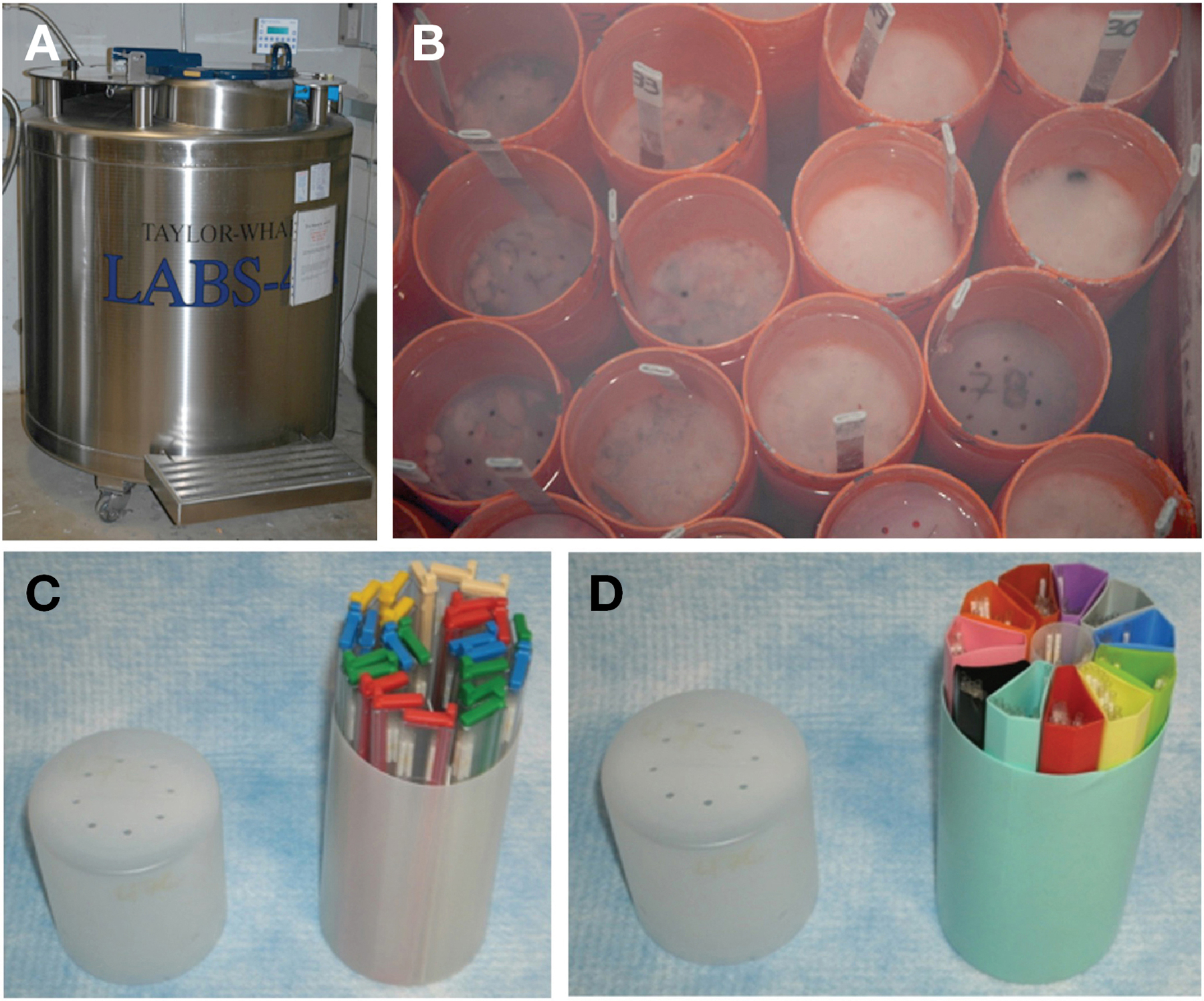
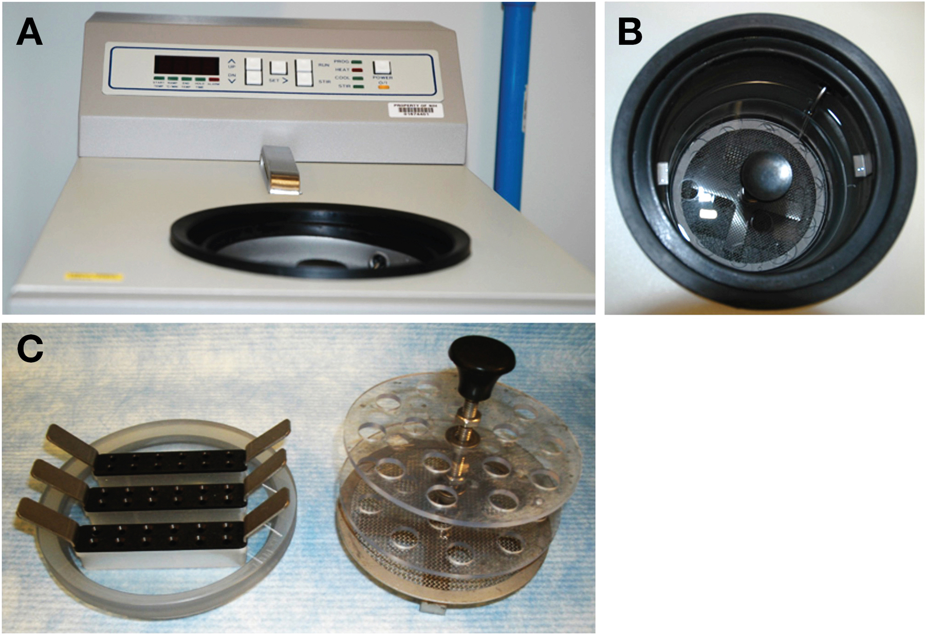
Protocols for cryopreservation of mouse embryos and sperm are important for preserving genetically engineered mice (GEMs) used in research to study human development and diseases. Embryo cryopreservation is mainly carried out using either of two protocols: controlled gradual cooling or vitrification. Sperm cryopreservation protocols include two methodologies that are commonly referred to as JAX and CARD. Quality-control measures are necessary to ensure that GEMs are properly cryopreserved so that they can be retrieved for future use. An archiving system is also important in keeping proper records of frozen sperm and embryos. Frozen embryos and sperm are now preferred over live mice for shipping to distant locations. This article describes detailed protocols used in cryopreservation of mouse embryos and sperm, as well as their retrieval to live mice. © 2021 U.S. Government. Sperm cryopreservation Basic Protocol 1: JAX protocol for sperm cryopreservation Support Protocol 1: JAX protocol for making sperm cryopreservation medium Basic Protocol 2: JAX protocol for IVF of mouse sperm Alternate Protocol 1: Modified CARD protocol for sperm cryopreservation Support Protocol 2: CARD protocol for making sperm cryopreservation medium Alternate Protocol 2: CARD protocol for IVF of mouse sperm Embryo cryopreservation Basic Protocol 3: Cryopreserving and thawing 2-cell embryos Alternate Protocol 3: Cryopreserving and thawing 8-cell to morula-stage embryos Surgical transfer of embryos Basic Protocol 4: Infundibulum transfer of 2-cell to morula-stage embryos.
Keywords: In Vitro Fertilization (IVF); controlled rate freezing; embryo cryopreservation; sperm cryopreservation; vitrification.
Published 2021. This article is a U.S. Government work and is in the public domain in the USA.
Conflict of interest statement
Conflict of Interest
None
Figures

Similar articles
-
The CARD Method for Simple Vitrification of Mouse Oocytes: Advantages and Applications.Methods Mol Biol. 2019;1874:229-242. doi: 10.1007/978-1-4939-8831-0_13. Methods Mol Biol. 2019. PMID: 30353517
-
The CARD Method for Mouse Sperm Cryopreservation and In Vitro Fertilization Using Frozen-Thawed Sperm.Methods Mol Biol. 2019;1874:243-256. doi: 10.1007/978-1-4939-8831-0_14. Methods Mol Biol. 2019. PMID: 30353518
-
Applications of cryopreserved unfertilized mouse oocytes for in vitro fertilization.Cryobiology. 2013 Oct;67(2):188-92. doi: 10.1016/j.cryobiol.2013.06.011. Epub 2013 Jul 8. Cryobiology. 2013. PMID: 23846105
-
Optimized protocols for sperm cryopreservation and in vitro fertilization in the rat.Lab Anim (NY). 2022 Oct;51(10):256-274. doi: 10.1038/s41684-022-01053-5. Epub 2022 Oct 10. Lab Anim (NY). 2022. PMID: 36216983 Review.
-
Vitrification and conventional freezing methods in sperm cryopreservation: A systematic review and meta-analysis.Eur J Obstet Gynecol Reprod Biol. 2019 Feb;233:84-92. doi: 10.1016/j.ejogrb.2018.11.028. Epub 2018 Dec 14. Eur J Obstet Gynecol Reprod Biol. 2019. PMID: 30580229
Cited by
-
Mice on the move.Lab Anim (NY). 2021 Sep;50(9):233-235. doi: 10.1038/s41684-021-00829-5. Lab Anim (NY). 2021. PMID: 34373647 No abstract available.
References
Literature Cited
-
- Awasthi PR, French CF, Sztein J, Bedigian R, Sharp JJ, Lloyd KC 2003. Frozen sperm as an alternative to shipping live mice. Contemporary Topics in Laboratory Animal Science. 42: 8–11. - PubMed
Key References:
-
- Ostermeier et al., 2008. Is the original reference for the Jackson Laboratory method of mouse sperm cryopreservation. See above
-
- Renard and Babinet 1984. Is a key reference on the mouse embryo cryopreservation method. It is the basis of what a lot of labs are using to freeze down mouse embryos. See above
-
- Hogan et al., 1994. Good reference overall reference on the surgical transfer of embryos. See above
-
- Longenecker and Kulkarni, 2009. Good reference on trouble shooting of mouse surgeries. See above
Internet Resources:
Protocols and additional information
-
- www.transtechsociety.org.
-
This is the website for the International Society for Transgenic Technologies (ISTT). It offers information related to items described in this manuscript, but also provides information on a variety of topics related to the field of transgenic technology
-
- http://card.medic.kumamoto-u.ac.jp/card/english/sigen/manual/onlinemanua....
-
This is the website for CARD related techniques and methods.
-
- www.jax.org.
-
The Jackson Laboratory provides workshops in the cryopreservation of mouse embryos and sperm.
-
- www.infrafrontier.eu.
-
(INFRAFRONTIER mouse disease models) This website is related to the European Mouse Mutant Archive (EMMA) and is another useful website in mouse cryopreservation protocols and other useful resources. Genetically engineered mice can also be found here.
Finding genetically modified mice
-
- www.findmice.org.
-
International Mouse Strain Resource (IMSR). This is an important resource for finding various mouse models that are available from various repositories IMSR
-
- www.mmrc.org.
-
This the website for the Mutant Mouse Resource & Research Centers (MMRC). This a website supported by the NIH. It is another useful resource in finding previously genetically engineered mice.
-
- www.informatics.jax.org.
-
Mouse Genome Informatics (MGI) Another website useful relating to genetically modified mice.
-
- https://mus.brc.riken.jp/en/
-
RIKENBRC Experimental Mouse Division
Shipping of Biological Samples
-
- www.saftpak.com.
-
Helpful with the shipment of biological samples using liquid nitrogen and dry ice. Company offers courses in the safe transport of infectious substances, biological samples, dry ice, liquid nitrogen, and related materials.
-
- www.aphis.usda.gov/aphis/ourfocus/animalhealth/export.
-
Website of the Animal Plant and Health Inspection Service of the USDA. Information available on information of importation and exportation of animal related products.
MeSH terms
Grants and funding
LinkOut - more resources
Full Text Sources
Other Literature Sources
