

Dome-shaped macula in premature infants visualized by handheld spectral-domain optical coherence tomography
- PMID: 34044111
- PMCID: PMC8328941
- DOI: 10.1016/j.jaapos.2020.12.007
Dome-shaped macula in premature infants visualized by handheld spectral-domain optical coherence tomography
Abstract
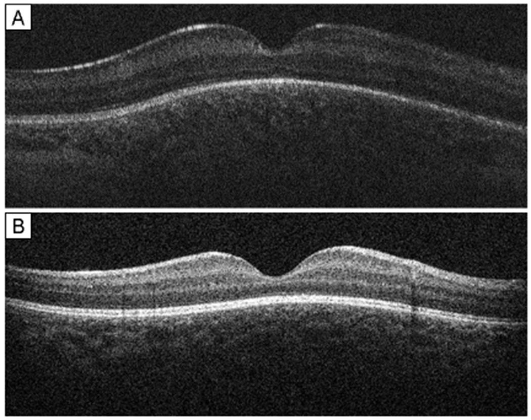
Purpose: To describe dome-shaped macula and associated clinical findings in premature infants.
Methods: This prospective, observational cohort study included a consecutive sample of premature infants screened for retinopathy of prematurity (ROP) with 9-month follow-up. Handheld spectral domain optical coherence tomography (SD-OCT) was performed at the time of ROP screening. Images were assessed for dome-shaped macula, cystoid macular edema, epiretinal membrane, vitreous bands, and punctate hyperreflective vitreous opacities. Dome height measurements were performed in a subset of images. Teller visual acuity and cycloplegic refraction were performed at an adjusted age of 8-10 months.
Results: Of 37 infants (74 eyes; 49% male; mean gestational age 27.8 ± 3.2 weeks; mean birth weight 949 ± 284 g), 24/37 (65%) demonstrated dome-shaped macula in at least one eye (13 both eyes, 5 right eye only, and 6 left eye only). Of the 74 eyes, 26 (35%) could be reliably measured, with a mean dome height of 139.0 ± 72.3 μm (range, 54-369 μm). Presence of dome-shaped macula was associated with a diagnosis of ROP (P = 0.02; OR, 3.03; 95% CI, 1.18-7.82) and pre-plus or plus disease (P = 0.02; OR, 4.20; 95% CI, 1.05-16.78). Infants with dome-shaped macula had lower birth weight compared with those without (877 vs 1081 g; P = 0.04). No associations with other demographics, OCT findings, and 9-month refractive outcomes were found.
Conclusions: Dome-shaped macula was frequently identified by handheld SD-OCT in premature infants, especially those with lower birth weight and severe ROP. The long-term clinical significance of this finding is unknown.
Copyright © 2021 American Association for Pediatric Ophthalmology and Strabismus. Published by Elsevier Inc. All rights reserved.
Figures
References
-
- Fielder A, Blencowe H, O’Connor A, Gilbert C. Impact of retinopathy of prematurity on ocular structures and visual functions. Arch Dis Child Fetal Neonatal Ed 2015;100:F179–84. - PubMed
Publication types
MeSH terms
Grants and funding
LinkOut - more resources
Full Text Sources
Other Literature Sources
