

Branching development of early post-implantation human embryonic-like tissues in 3D stem cell culture
- PMID: 34044259
- PMCID: PMC8325636
- DOI: 10.1016/j.biomaterials.2021.120898
Branching development of early post-implantation human embryonic-like tissues in 3D stem cell culture
Abstract
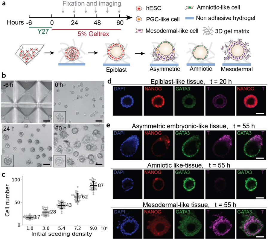
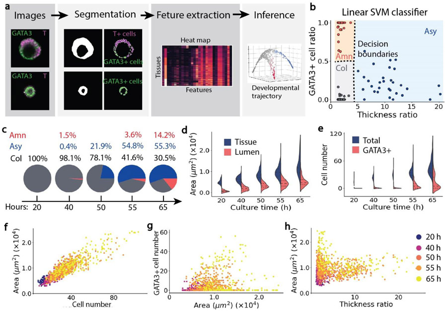
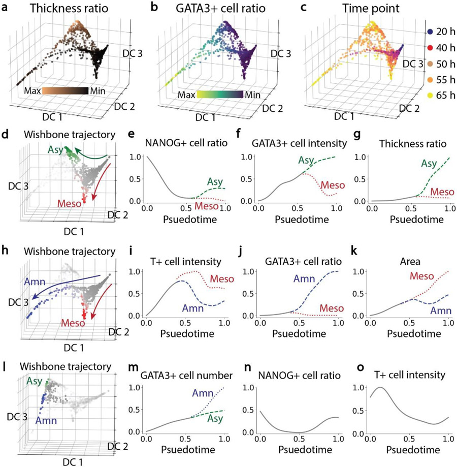
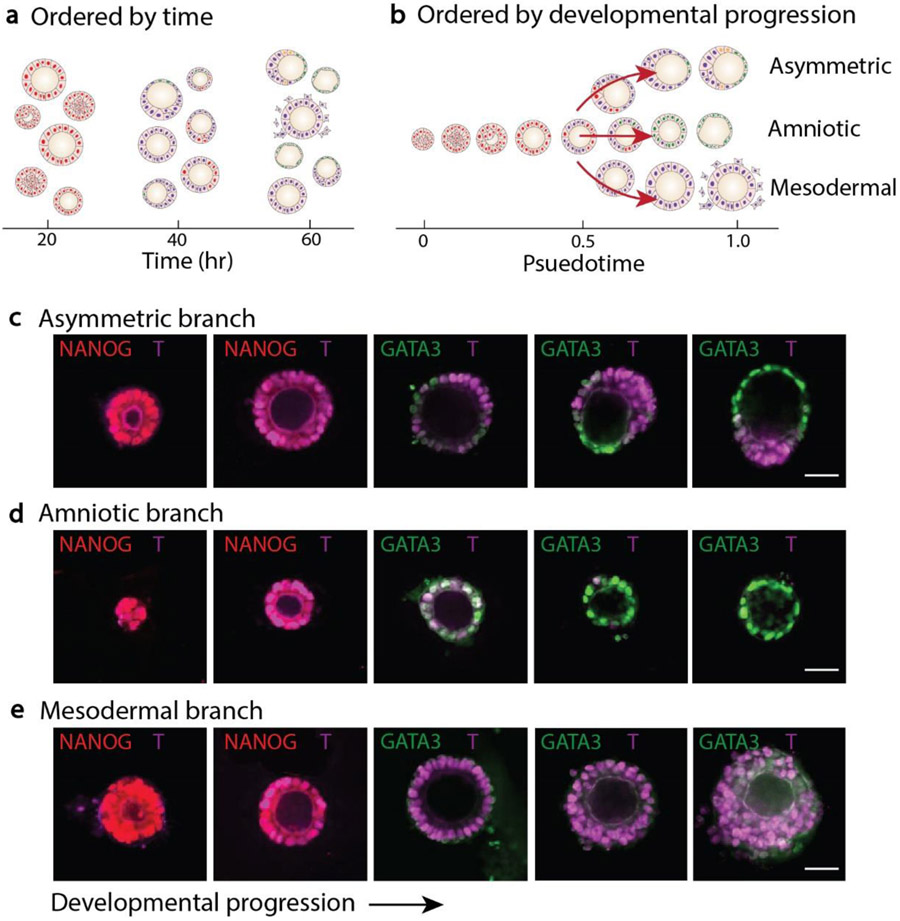
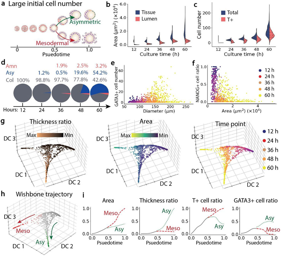
Human embryonic stem cells (hESCs) have the intrinsic capacity to self-organize and generate patterned tissues. In vitro models that coax hESCs to form embryonic-like structures by modulating physical environments and priming with chemical signals have become a powerful tool for dissecting the regulatory mechanisms underlying early human development. Here we present a 3D suspension culture system of hESCs that can generate post-implantation, pre-gastrulation embryonic-like tissues in an efficient and controllable manner. The efficiency of the development of asymmetric tissues, which mimic the post-implantation, pre-gastrulation amniotic sac, was about 50% in the 3D suspension culture. Quantitative imaging profiling and unsupervised trajectory analysis revealed that hESC aggregates first entered into a transitional stage expressing Brachyury (or T), before their development branched into different paths to develop into asymmetric embryonic-like tissues, amniotic-like tissues, and mesodermal-like tissues, respectively. Moreover, the branching developmental trajectory of embryonic-like structures was affected by the initial cell seeding density or cluster size of hESCs. A higher percentage of amniotic-like tissues was observed under a small initial cell seeding density of hESCs. Conversely, a large initial cell seeding density of hESCs promoted the development of mesodermal-like tissues. Intermediate cell seeding densities of hESCs in the 3D suspension culture promoted the development of asymmetric embryonic-like tissues. Our results suggest that hESCs have the intrinsic capability to sense the initial cell population size, which in turn regulates their differentiation and self-organization into different embryonic-like tissues. Our 3D suspension culture thus provides a promising experimental tool to study the interplay between tissue topology and self-organization and progressive embryonic development using in vitro hESC-based models.
Keywords: Branching tissue development; Human pluripotent stem cells; Synthetic embryonic model.
Copyright © 2021 Elsevier Ltd. All rights reserved.
Conflict of interest statement
Declaration of interests
The authors declare that they have no known competing financial interests or personal relationships that could have appeared to influence the work reported in this paper.
Figures

Similar articles
-
Development of a 48-Well Dynamic Suspension Culture System for Pancreatic Differentiation from Human Embryonic Stem Cells.Stem Cell Rev Rep. 2022 Apr;18(4):1423-1433. doi: 10.1007/s12015-021-10312-w. Epub 2021 Dec 2. Stem Cell Rev Rep. 2022. PMID: 34855111
-
Large-scale generation of megakaryocytes from human embryonic stem cells using transgene-free and stepwise defined suspension culture conditions.Cell Prolif. 2021 Apr;54(4):e13002. doi: 10.1111/cpr.13002. Epub 2021 Feb 21. Cell Prolif. 2021. PMID: 33615584 Free PMC article.
-
In vitro neural differentiation of human embryonic stem cells using a low-density mouse embryonic fibroblast feeder protocol.Methods Mol Biol. 2010;584:71-95. doi: 10.1007/978-1-60761-369-5_4. Methods Mol Biol. 2010. PMID: 19907972
-
Signaling mechanisms that direct cell fate specification and morphogenesis in human embryonic stem cells-based models of human gastrulation.Emerg Top Life Sci. 2023 Dec 18;7(4):383-396. doi: 10.1042/ETLS20230084. Emerg Top Life Sci. 2023. PMID: 38087898 Review.
-
The tumorigenicity of human embryonic stem cells.Adv Cancer Res. 2008;100:133-58. doi: 10.1016/S0065-230X(08)00005-5. Adv Cancer Res. 2008. PMID: 18620095 Review.
Cited by
-
Deep manifold learning reveals hidden developmental dynamics of a human embryo model.Sci Adv. 2025 Aug 8;11(32):eadr8901. doi: 10.1126/sciadv.adr8901. Epub 2025 Aug 6. Sci Adv. 2025. PMID: 40768579 Free PMC article.
-
Temporally resolved early bone morphogenetic protein-driven transcriptional cascade during human amnion specification.Elife. 2024 Jul 25;12:RP89367. doi: 10.7554/eLife.89367. Elife. 2024. PMID: 39051990 Free PMC article.
-
Modeling development using hydrogels.Development. 2023 Jul 1;150(13):dev201527. doi: 10.1242/dev.201527. Epub 2023 Jun 30. Development. 2023. PMID: 37387575 Free PMC article.
-
Temporally resolved single cell transcriptomics in a human model of amniogenesis.bioRxiv [Preprint]. 2024 Aug 22:2023.09.07.556553. doi: 10.1101/2023.09.07.556553. bioRxiv. 2024. PMID: 39026707 Free PMC article. Preprint.
-
Machine learning-assisted imaging analysis of a human epiblast model.Integr Biol (Camb). 2021 Oct 15;13(9):221-229. doi: 10.1093/intbio/zyab014. Integr Biol (Camb). 2021. PMID: 34327532 Free PMC article.
References
-
- Harland Richard M. "A new view of embryo development and regeneration." Science 360, no. 6392 (2018): 967–968. - PubMed
-
- Shahbazi Marta N., and Zernicka-Goetz Magdalena. "Deconstructing and reconstructing the mouse and human early embryo." Nature cell biology 20, no. 8 (2018): 878–887. - PubMed
-
- Hyun Insoo, Wilkerson Amy, and Johnston Josephine. "Embryology policy: Revisit the 14-day rule." Nature 533, no. 7602 (2016): 169–171. - PubMed
-
- Pera Martin F. "Human embryo research and the 14-day rule." Development 144, no. 11 (2017): 1923–1925. - PubMed
Publication types
MeSH terms
Grants and funding
LinkOut - more resources
Full Text Sources
Other Literature Sources
