. 2021 Jan-Dec:30:9636897211019025.
doi: 10.1177/09636897211019025.

Immunosuppressant Drugs Mitigate Immune Responses Generated by Human Mesenchymal Stem Cells Transplanted into the Mouse Parenchyma

Affiliations

Immunosuppressant Drugs Mitigate Immune Responses Generated by Human Mesenchymal Stem Cells Transplanted into the Mouse Parenchyma

Jung Won Hwang et al. Cell Transplant. 2021 Jan-Dec.

Abstract

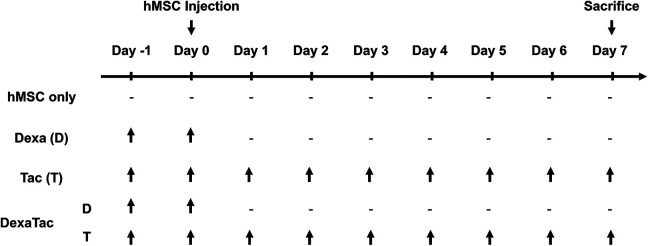
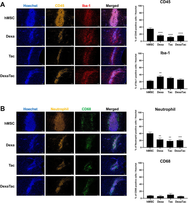
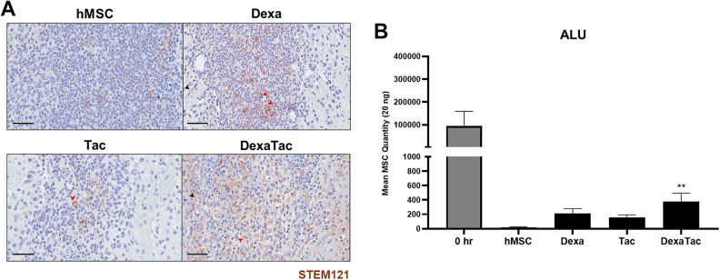
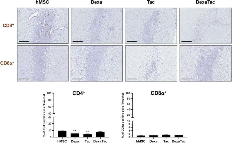
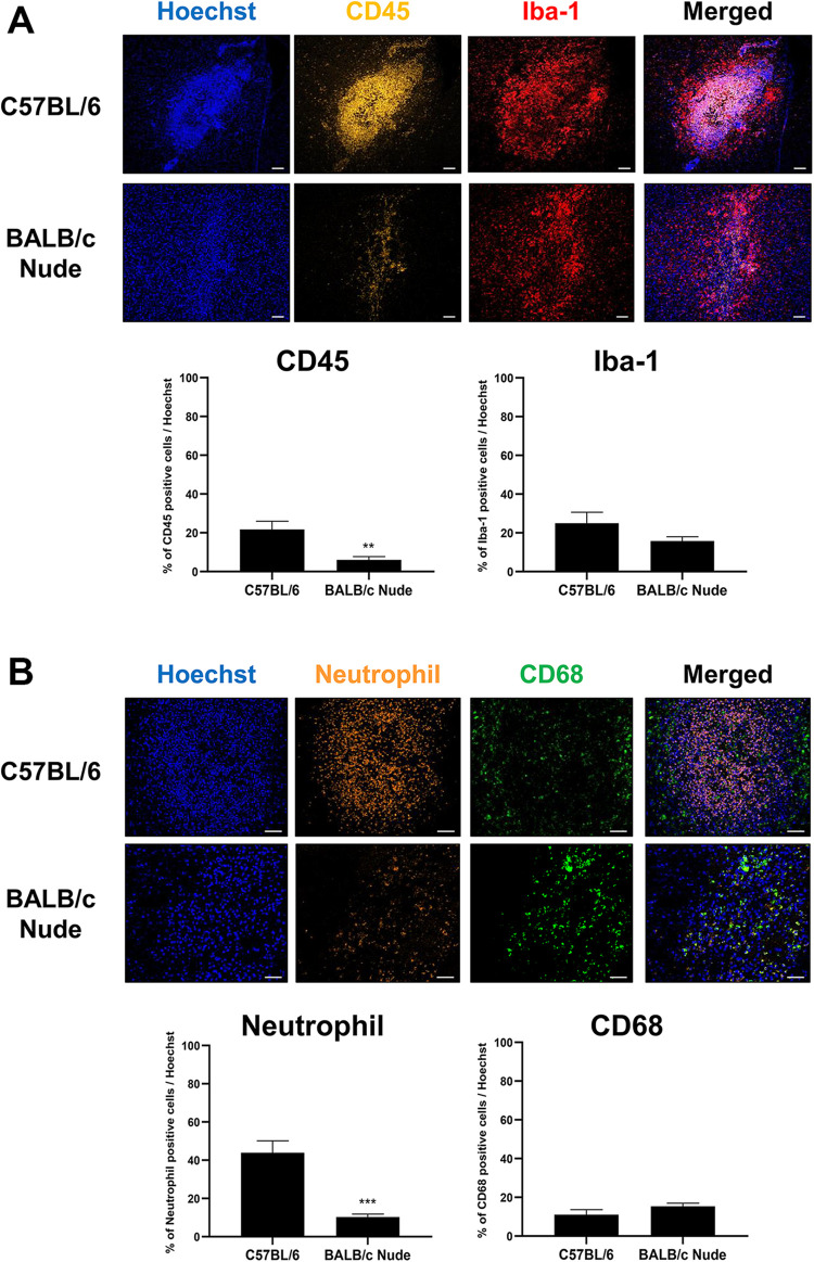
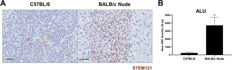
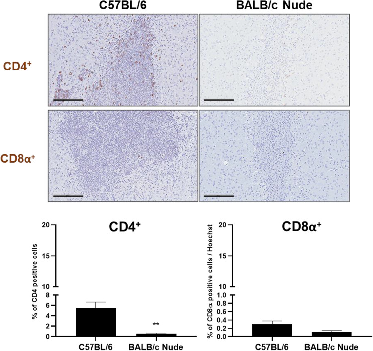
It has been widely accepted that mesenchymal stem cells (MSCs) can evade the immune surveillance of the recipient. However, emerging research cast doubt on whether MSCs are intrinsically immune-privileged. Previously, we observed that the transplantation of human MSCs (hMSCs) into the mouse parenchyma attracted a high infiltration of leukocytes into the injection tract. Thus, in order to reduce the immune responses generated by hMSCs, the aim of this study was to assess which immunosuppressant condition (dexamethasone only, tacrolimus only, or dexamethasone and tacrolimus together) would not only reduce the overall immune response but also enhance the persistence of MSCs engrafted into the caudate putamen of wild-type C57BL/6 mice. According to immunohistochemical analysis, compared to the hMSC only group, the administration of immunosuppressants (for all three conditions) reduced the infiltration of CD45-positive leukocytes and neutrophils at the site of injection. The highest hMSC persistence was detected from the group that received combinatorial administrations of dexamethasone and tacrolimus. Moreover, compared to the immunocompetent WT mouse, higher MSC engraftment was observed from the immunodeficient BALB/c mice. The results of this study support the use of immunosuppressants to tackle MSC-mediated immune responses and to possibly prolong the engraftment of transplanted MSCs.

Keywords: immunologic surveillance; immunosuppressive agents; mesenchymal stem cell; transplants.


Conflict of interest statement

Declaration of Conflicting Interests: The author(s) declared no potential conflicts of interest with respect to the research, authorship, and/or publication of this article.

Figures

Figure 1.
Figure 1.
Timeline of experiment. C57BL/6 mice are randomly allocated into four different groups: hMSC only, dexamethasone (Dexa; D) only, tacrolimus (Tac; T) only, and a combination of dexamethasone and tacrolimus (DexaTac). Human mesenchymal stem cells (hMSCs) are transplanted into the left CPu of wild-type C57BL/6 mice at Day 0. Mice are sacrificed at Day 7. Dexamethasone is administered at 1 mg/kg/day via oral gavage at Day -1 and Day 0. Tacrolimus is administered at 3 mg/kg/day via the intraperitoneal route daily starting from Day -1 up to the termination time point (Day 7). The DexaTac group is given administrations of both dexamethasone and tacrolimus via the respective administration routes and dosage. Note the hyphen indicates that no immunosuppressants have been administered.
Figure 2.
Figure 2.
Administration of immunosuppressants reduces the expression levels of CD45-positive leukocytes and neutrophils at the hMSC injection site. (A) Compared to the Dexa (n = 6), Tac (n = 5), and DexaTac (n = 7) groups, severe infiltration of CD45-positive leukocytes (indicated in golden yellow) is visualized from the injection site of the hMSC group (n = 7) via IHC staining. For all 4 groups (hMSC, Dexa, Tac, and DexaTac), Iba-1 microglia cells (indicated in red) are randomly dispersed at the injection site and the region surrounding the cell graft. (B) The hMSC group shows the highest infiltration of neutrophils (indicated in golden yellow) at the injection site. The expression of neutrophils is significantly reduced for the Dexa (n = 6), Tac (n = 5), and DexaTac (n = 7) groups. CD68-positive macrophages (indicated in green) are not as densely populated as neutrophils. Macrophages are identified at and the region bordering the engraftment site. Statistical significance is defined as ** P < 0.01, *** P < 0.001, **** P < 0.0001 vs. hMSC; mean ± SEM (One-way ANOVA). Scale bar = 100 µm.
Figure 3.
Figure 3.
Residual hMSCs remaining in the mouse parenchyma following immunosuppressant administration at Day 7. (A) IHC results of representative tissue sections from the hMSC (n = 7), Dexa (n = 6), Tac (n = 5), and DexaTac (n = 7) groups, respectively, show STEM121 positive cells that are discernible as brown signals surrounding the nuclei of the cells (solid red arrowhead). Non-specific brown signals, not enclosing the nuclei of cells, can also be identified in the surrounding tissue regions of the injection site (solid black arrowhead). (B) The number of residual hMSCs present in the parenchyma of the 0 hr (n = 3), hMSC (n = 6), Dexa (n = 7), Tac (n = 6), and DexaTac (n = 7) groups, respectively, is quantitated via ALU qPCR. 0 hr is the positive control group where unlike the other experimental groups, this group was sacrificed not at Day 7 but immediately following transplantation of hMSCs. Out of the immunosuppressant groups, the highest number of persisting hMSCs is observed from the DexaTac group. Statistical significance is defined as ** P < 0.01 vs. hMSC; mean ± SEM (One-way ANOVA). Scale bar = 50 µm.
Figure 4.
Figure 4.
Administering immunosuppressants decreases the proliferation of CD4+ T cells at the hMSC engraftment site. The expressions of CD4+ (top row) and CD8α+ (bottom row) T cells at the site of hMSC injection are assessed via IHC staining for the hMSC (n = 7), Dexa (n = 6), Tac (n = 5), and DexaTac (n = 7) groups, respectively. T cells are identified as dark brown signals colocalized to the nuclei of cells. CD4+ T cells are sparsely distributed and CD8α+ T cells are barely identified at the site where hMSCs have been implanted. Statistical significance is defined as ** P < 0.01 vs. hMSC; mean ± SEM (One-way ANOVA). Scale bar = 200 µm.
Figure 5.
Figure 5.
Infiltration of leukocytes and neutrophils are attenuated at the injection site of BALB/c nude mice. (A) Based on IHC staining, the percentage of CD45-positive leukocytes (indicated in golden yellow) is greatly reduced in the injection site of BALB/c nude mice (n = 7) compared to that of C57BL/6 mice (n = 4). Notable and significant differences are not observed when comparing the percentages of Iba-1 positive microglia cells (indicated in red) from both groups. (B) Contrary to C57BL/6 mice (n = 4), severe infiltration of neutrophils (indicated in orange) is not identified from the injection site of BALB/c nude mice (n = 7). The percentage of CD68-positive macrophages was slightly higher in the C57BL/6 group in comparison to that of BALB/c nude mice but there was no significant difference between the groups. Statistical significance is defined as **P < 0.01, *** P < 0.001 vs. C57BL/6 mice; mean ± SEM (t-test). Scale bars: (A) 100 µm, (B) 50 µm.
Figure 6.
Figure 6.
Higher number of residual hMSCs is quantitated from the parenchyma of BALB/c nude mice at Day 7. (A) Via IHC staining, STEM121-positive cells are identified as brown signals from representative tissue sections of both C57BL/6 (n = 5) and BALB/c nude mice (n = 5). (B) The number of residual hMSCs persisting in the parenchyma of both C57BL/6 (n = 5) and BALB/c nude mice (n = 5) is assessed via ALU qPCR. MSC persistence is higher in BALB/c nude mice. Statistical significance is defined as **P < 0.01 vs. C57BL/6 mice; mean ± SEM (t-test). Scale bar = 50 µm.
Figure 7.
Figure 7.
T cell reactivity is attenuated at the hMSC engraftment site of BALB/c nude mice. The proliferation of CD4+ (top row) and CD8α+ (bottom row) T cells at the site of hMSC injection in C57BL/6 (n = 4) and BALB/c nude mice (n = 7) is evaluated via IHC staining. Dark brown signals, indicating the presence of T cells, can be clearly identified from the representative tissue section of C57BL/6 mice stained with the CD4 antibody while positive signals are barely detectable from representative tissue sections of BALB/c nude mice. Statistical significance is defined as **P < 0.01 vs. C57BL/6 mice; mean ± SEM (t-test). Scale bar = 200 µm.

References

    1. Han Y, Li X, Zhang Y, Han Y, Chang F, Ding J. Mesenchymal stem cells for regenerative medicine. Cells. 2019;8(8):886. - PMC - PubMed
    1. Parekkadan B, Milwid JM. Mesenchymal stem cells as therapeutics. Annu Rev Biomed Eng. 2010;12:87–117. - PMC - PubMed
    1. Caplan AI, Correa D. The MSC: an injury drugstore. Cell Stem Cell. 2011;9(1):11–15. - PMC - PubMed
    1. Baraniak PR, McDevitt TC. Stem cell paracrine actions and tissue regeneration. Regen Med. 2010;5(1):121–143. - PMC - PubMed
    1. Park SE, Lee NK, Lee J, Hwang JW, Choi SJ, Hwang H, Hyung B, Chang JW, Na DL. Distribution of human umbilical cord blood-derived mesenchymal stem cells in the Alzheimer’s disease transgenic mouse after a single intravenous injection. Neuroreport. 2016;27(4):235–241. - PubMed

Publication types

MeSH terms

Substances

LinkOut - more resources