Review
. 2021 Sep;32(9):2125-2136.
doi: 10.1681/ASN.2021010042. Epub 2021 May 27.

Molecular Mechanisms of Renal Magnesium Reabsorption

Affiliations
Review

Molecular Mechanisms of Renal Magnesium Reabsorption

David H Ellison et al. J Am Soc Nephrol. 2021 Sep.

Abstract

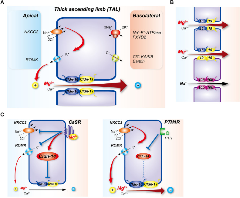
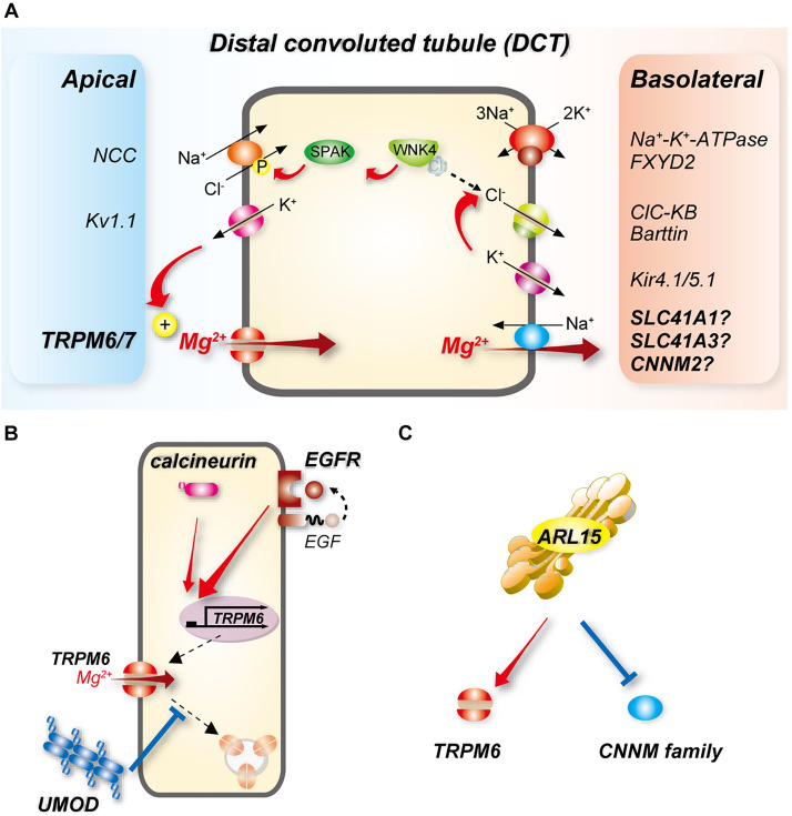
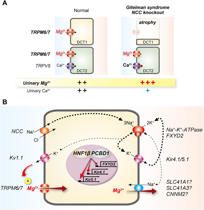
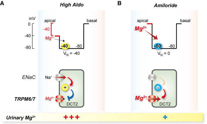
Magnesium is an essential cofactor in many cellular processes, and aberrations in magnesium homeostasis can have life-threatening consequences. The kidney plays a central role in maintaining serum magnesium within a narrow range (0.70-1.10 mmol/L). Along the proximal tubule and thick ascending limb, magnesium reabsorption occurs via paracellular pathways. Members of the claudin family form the magnesium pores in these segments, and also regulate magnesium reabsorption by adjusting the transepithelial voltage that drives it. Along the distal convoluted tubule transcellular reabsorption via heteromeric TRPM6/7 channels predominates, although paracellular reabsorption may also occur. In this segment, the NaCl cotransporter plays a critical role in determining transcellular magnesium reabsorption. Although the general machinery involved in renal magnesium reabsorption has been identified by studying genetic forms of magnesium imbalance, the mechanisms regulating it are poorly understood. This review discusses pathways of renal magnesium reabsorption by different segments of the nephron, emphasizing newer findings that provide insight into regulatory process, and outlining critical unanswered questions.

Keywords: Gitelman syndrome; cell and transport physiology; claudin; ion transport; magnesium.


Figures

Figure 1.
Figure 1.
Mg2+ reabsorption along the TAL. (A) Mg2+ pathways and regulation. The basolateral Na+-K+-ATPase with its regulatory γ-subunit FXYD2 provides the drive for transcellular entry of Na+, K+, and Cl through the NKCC2. Recycling of K+ through ROMK contributes to the generation of the transepithelial voltage that provides the drive for paracellular movement of Mg2+ and Ca2+ through the claudin-16/19 complex (Cldn-16/19). Claudin-14 (Cldn-14) inhibits movement through Cldn-16/19, and its expression is stimulated by Mg2+ activation of CaSR or repressed by activation of the PTH1 receptor (PTH1R), through mechanisms that involve microRNAs. Chloride exits at the basolateral membrane via CLC-K channels. (B) Recent data suggest TAL expression of claudins displays a mosaic pattern, suggesting greater complexity than shown in (A). Proposed scheme for pore composition is on the basis of immunolocalization and single-cell RNA sequencing. Cells outlined in blue express claudins 16 (blue) and 19 (yellow), whereas those outlined in purple express claudins 10b (purple) and 19 but not claudin-16. Claudins form dimers within the same cell (cis interactions) and interact with dimers in adjacent cells to form ion pores (trans interactions). Adjacent cells expressing claudin 16 and 19 heterodimers form a Ca2+/Mg2+ pore. Cells expressing claudin-10b homodimers form an Na+ pore, which may regulate paracellular Ca2+/Mg2+ flux by modulating the transepithelial voltage. In claudin-10b–expressing cells, claudin-19 homodimers may form a Ca2+/Mg2+ pore with claudin-16/19 heterodimers in an adjacent cell. (C) Claudin-14 inhibits Ca2+/Mg2+ movement through Cldn-16/19, and its expression is stimulated by Mg2+ activation of CaSR (left), or inhibited by activation of PTH1R (right). CaSR also exerts inhibitory effects on NKCC2 and ROMK (left).
Figure 2.
Figure 2.
Transcellular Mg2+ reabsorption along the DCT. (A) Mg2+ enters cells along the early (DCT1) and late DCT (DCT2) through TRPM6, which requires interaction with TRPM7 for maximum activity. The basolateral exit pathway remains to be identified, but candidates are shown. Because transport through NCC is electroneutral, the potassium channel Kv1.1 is believed to generate the drive for Mg2+ entry at the apical membrane. NCC is activated by phosphorylation via the WNK-SPAK kinase pathway. WNK kinases are inhibited by direct binding of Cl; coupling of K+ and Cl efflux via Kir4.1/5.1 and CLC-KB as a result of increased plasma [K+] lowers intracellular [Cl] leading to WNK kinase activation. Energy consumption by the basolateral Na+-K+-ATPase drives all of these processes (see Figure 3). (B) Activation of EGFR induces TRPM6 mRNA expression and activity, and calcineurin may promote this. UMOD may stimulate TRPM6 activity by inhibiting its endocytosis. (C) ARL15, a GTP-binding protein, may activate TRPM6 but inhibit cyclin and CNNMs.
Figure 3.
Figure 3.
Links between activities of NCC and the Na+-K+-ATPase and DCT Mg2+ reabsorption. (A) DCT1 atrophy occurs with disruption of NCC activity, and is seen in Gitelman syndrome (GS, NCC mutation) and in NCC knockout mice; and in epilepsy, ataxia, sensorineural deafness, and tubulopathy (EAST) syndrome (Kir4.1 mutation); Bartter syndrome types 3 and 4 (CLC-KB and barttin mutations); and Kir4.1 knockout mice (not shown). This reduces the capacity for Mg2+ reabsorption in DCT1, leading to hypomagnesemia. The DCT2 does not compensate for this effect, and Mg2+ reabsorption may also be lower in this segment due to NCC inhibition. Calcium (Ca2+) is not reabsorbed along the DCT1, but along the DCT2 and connecting segment (not shown). In GS, Ca2+ reabsorption is paradoxically enhanced leading to hypocalciuria; several mechanisms have been proposed, including enhanced entry along the DCT2, although the evidence better supports increased Ca2+ PT reabsorption. (B) Generation of the driving force for Mg2+ reabsorption requires activity of the basolateral Na+-K+-ATPase. Consistent with this, mutations in its γ-subunit (FXYD2, shown in brown) cause renal Mg2+ wasting. The Na+-K+-ATPase maintains intracellular K+; apical efflux of K+ through Kv1.1 may determine the membrane potential for apical Mg2+ entry. Na+-K+-ATPase–mediated generation of an Na+ gradient at the basolateral membrane may provide the driving force for Mg2+ exit through an Na+-Mg2+ exchanger. Apical entry of Na+ via the NCC also contributes to maintenance of Na+-K+-ATPase activity. Thicker arrows represent established pathways for ion movement, whereas thinner arrows present proposed pathways. The transcription factor HNF1β and its dimerization cofactor PCBD1 both regulate expression of FXYD2, and when mutated cause hypomagnesemia. HNF1β has also been shown to regulate expression of Kir4.1 and Kir5.1.
Figure 4.
Figure 4.
Proposed mechanism of amiloride inhibition of Mg2+ wasting along the late distal convoluted tubule (DCT2). (A) In states of high aldosterone (High Aldo) the apical membrane is depolarized as a result of sodium entry via the ENaC (top panel shows voltage trace). This means that the transepithelial voltage is oriented with the lumen negative, relative to the basal side. The depolarized apical membrane voltage drives little Mg2+ into the cell through TRPM6/7, as the membrane voltage across the apical membrane is the primary driving force. The bottom panel shows this schematically. (B) Amiloride treatment inhibits sodium entry through ENaC, eliminating the negative transepithelial potential and hyperpolarizing the apical membrane (top panel shows voltage trace), leading to a net increase in the driving force for Mg2+ to enter the cell across the apical membrane through TRPM6/7. The bottom panel shows this schematically. VTE, transepithelial voltage.

References

    1. Jahnen-Dechent W, Ketteler M: Magnesium basics. Clin Kidney J 5: i3–i14, 2012 - PMC - PubMed
    1. de Baaij JH, Hoenderop JG, Bindels RJ: Magnesium in man: implications for health and disease. Physiol Rev 95: 1–46, 2015 - PubMed
    1. Wong NL, Dirks JH, Quamme GA: Tubular reabsorptive capacity for magnesium in the dog kidney. Am J Physiol 244: F78–F83, 1983 - PubMed
    1. Wong NL, Whiting SJ, Mizgala CL, Quamme GA: Electrolyte handling by the superficial nephron of the rabbit. Am J Physiol 250: F590–F595, 1986 - PubMed
    1. Brunette MG, Vigneault N, Carriere S: Micropuncture study of magnesium transport along the nephron in the young rat. Am J Physiol 227: 891–896, 1974 - PubMed

MeSH terms

LinkOut - more resources