. 2021 May 27;12(1):3201.
doi: 10.1038/s41467-021-23443-y.

Exploring protein hotspots by optimized fragment pharmacophores

Affiliations

Exploring protein hotspots by optimized fragment pharmacophores

Dávid Bajusz et al. Nat Commun. .

Abstract

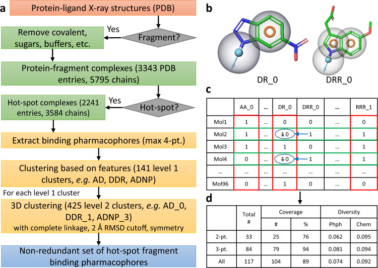
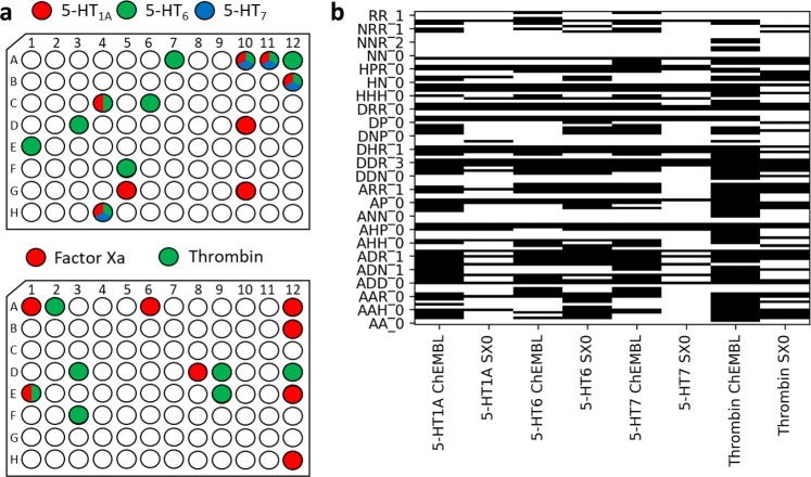
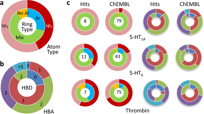
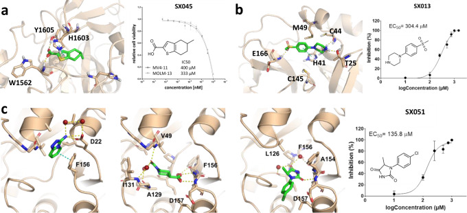
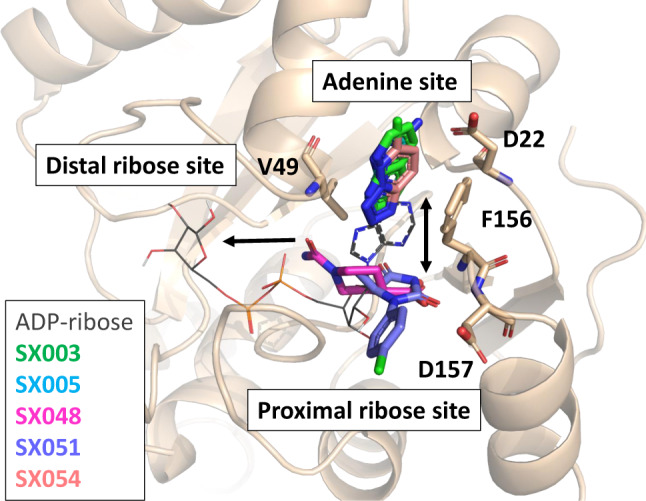
Fragment-based drug design has introduced a bottom-up process for drug development, with improved sampling of chemical space and increased effectiveness in early drug discovery. Here, we combine the use of pharmacophores, the most general concept of representing drug-target interactions with the theory of protein hotspots, to develop a design protocol for fragment libraries. The SpotXplorer approach compiles small fragment libraries that maximize the coverage of experimentally confirmed binding pharmacophores at the most preferred hotspots. The efficiency of this approach is demonstrated with a pilot library of 96 fragment-sized compounds (SpotXplorer0) that is validated on popular target classes and emerging drug targets. Biochemical screening against a set of GPCRs and proteases retrieves compounds containing an average of 70% of known pharmacophores for these targets. More importantly, SpotXplorer0 screening identifies confirmed hits against recently established challenging targets such as the histone methyltransferase SETD2, the main protease (3CLPro) and the NSP3 macrodomain of SARS-CoV-2.


Conflict of interest statement

Péter Pallai is the CEO and owner of Bioblocks. All remaining authors declare no competing interests.

Figures

Fig. 1
Fig. 1. SpotXplorer workflow.
a Computational workflow for assembling the non-redundant set of fragment binding pharmacophores. Fragment-sized ligands from the Protein Data Bank (PDB) were subjected to large-scale FTMap, analysis to identify hotspot binding fragments. Pharmacophore features (A—H-bond acceptor, D—H-bond donor, H—hydrophobic group, N—negative charge, P—positive charge, R—aromatic ring, see Supplementary Information, section 1.7 for more detail) with the largest contributions to the overall free energy of binding were clustered based on their respective feature sets and binned by paired root-mean-square deviation (RMSD) values (e.g., DRR_0, with 0 being an arbitrarily assigned identifier). b Pharmacophore models overlaid onto the co-crystallized representative ligand of the cluster. For each fragment fingerprint generated, fit to a larger model necessarily includes smaller models. Here, DR_0 can be fitted onto the corresponding features of DRR_0; the former is a submodel of the latter. c Compound selection algorithm. Iterative minimization of both the mean pairwise fingerprint similarity (green rows) and pharmacophore similarity (red columns) with submodels set to zero and simultaneous maximization of the total number of represented pharmacophores generated the SpotXplorer0 pilot library (96 compounds). d Final coverage and distribution data of the 96-compound set selected using 2-point and 3-point pharmacophores (along with the total numbers of non-redundant 2-point and 3-point pharmacophores). Pharmacophore (Phph) and chemical (Chem) diversity is measured as mean Tanimoto similarities.
Fig. 2
Fig. 2. Experimental validation of SpotXplorer0 on known target classes.
a SpotXplorer0 pilot library screening yielded a diverse set of confirmed hits for validated GPCR and protease drug targets. Hits are denoted by color in their respective plate positions (upper panel: 5-HT1A with red, 5-HT6 with green, 5-HT7 with blue, lower panel: factor Xa with red, thrombin with green). b The handful of hit compounds from SpotXplorer0 represents the majority of pharmacophore models (black) that occur over the respective sets of (typically 4–11 times more) fragment-sized ligands in the ChEMBL database (Percentages are reported in Table 1).
Fig. 3
Fig. 3. Topological features of SpotXplorer0 hits vs. ChEMBL fragments.
a Selection by pharmacophore results in a relatively even distribution of both ring types (Ar: only aromatic—light blue, Mix: both aromatic and non-aromatic—light green, No Ar: non-aromatic—yellow) and both atom types (sp2 hybridization—dark red, vs. sp3 hybridization—light red/salmon). b Overall hydrogen bond distribution of ligands in SpotXplorer0 resulting from pharmacophore matching (HBD: no. of H-bond donors, HBA: no. of H-bond acceptors; 0—dark blue, 1—red, 2—light green, 3—purple, >3—light blue). c Comparison of SpotXplorer0 hit compounds and ChEMBL fragments for selected targets. Each of the targets shown selects non-overlapping ring subsets: 5-HT1A: mixed ring types with high sp3 character and few hydrogen bond sites; 5-HT6: flatter rings, more aromatic character with moderate hydrogen bond site counts; Thrombin: non-aromatic sp2 rings with higher hydrogen bond site counts (Colors are identical to panels a and b.).
Fig. 4
Fig. 4. Binding poses and cellular activities of SpotXplorer0 hits against challenging and current targets.
a Predicted binding pose of the fragment SX045 (green) in the binding pocket of the SETD2 histone methyltransferase. The fragment decreases the viability of MV4-11 and MOLM-13 leukemia cells in a dose-dependent manner (see Supplementary Information, section 8), with IC50 values of 400 µM and 333 µM, respectively. b X-ray structure of the fragment SX013 in complex with the main protease (3CLPro) of the SARS-CoV-2 virus (PDB entry 5RHD). The fragment inhibits the SARS-COV-2-induced mortality of Vero E6 cells with an EC50 of 304 µM. c X-ray structures of fragments SX005, SX048 and SX051 (left to right) in complex with the NSP3 macrodomain of the SARS-CoV-2 virus (PDB entries 5S4G, 5S4H, and 5S4I). The fragments show antiviral activities with EC50 values in the high micromolar range in the cellular assay. (In the IC50 and EC50 plots, data are presented as mean values +/− SD, calculated from n = 3 biologically independent samples.) Source data are provided in the Source Data file.
Fig. 5
Fig. 5. SpotXplorer0 hits against the SARS-CoV-2 NSP3 macrodomain.
Overlay of the binding poses of the five SpotXplorer0 hits against the SARS-CoV-2 NSP3 macrodomain (colored sticks, SX003—green, SX005—light blue, SX048—magenta, SX051—purple, SX054—orange) with the binding pose of ADP-ribose (gray lines, from PDB structure 6WOJ). Three and two hits occupy the adenine and proximal ribose sites, respectively, providing merging and growing options towards the neighboring subsites (indicated by black arrows).

Similar articles

Cited by

References

    1. Williams G, Ferenczy GG, Ulander J, Keserű GM. Binding thermodynamics discriminates fragments from druglike compounds: a thermodynamic description of fragment-based drug discovery. Drug Discov. Today. 2017;22:681–689. doi: 10.1016/j.drudis.2016.11.019. - DOI - PubMed
    1. Ferenczy GG, Keserű GM. Thermodynamic profiling for fragment-based lead discovery and optimization. Expert Opin. Drug Discov. 2020;15:117–129. doi: 10.1080/17460441.2020.1691166. - DOI - PubMed
    1. Kozakov D, et al. Ligand deconstruction: Why some fragment binding positions are conserved and others are not. Proc. Natl Acad. Sci. 2015;112:E2585–E2594. doi: 10.1073/pnas.1501567112. - DOI - PMC - PubMed
    1. Murray CW, Verdonk ML. The consequences of translational and rotational entropy lost by small molecules on binding to proteins. J. Comput. Aided Mol. Des. 2002;16:741–753. doi: 10.1023/A:1022446720849. - DOI - PubMed
    1. Ferenczy GG, Keserű GM. Thermodynamics of fragment binding. J. Chem. Inf. Model. 2012;52:1039–1045. doi: 10.1021/ci200608b. - DOI - PubMed

Publication types

MeSH terms

Substances

LinkOut - more resources