

Metabolomics study of fibroblasts damaged by UVB and BaP
- PMID: 34045475
- PMCID: PMC8160258
- DOI: 10.1038/s41598-021-90186-7
Metabolomics study of fibroblasts damaged by UVB and BaP
Abstract
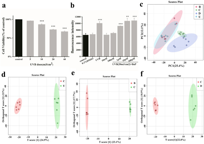
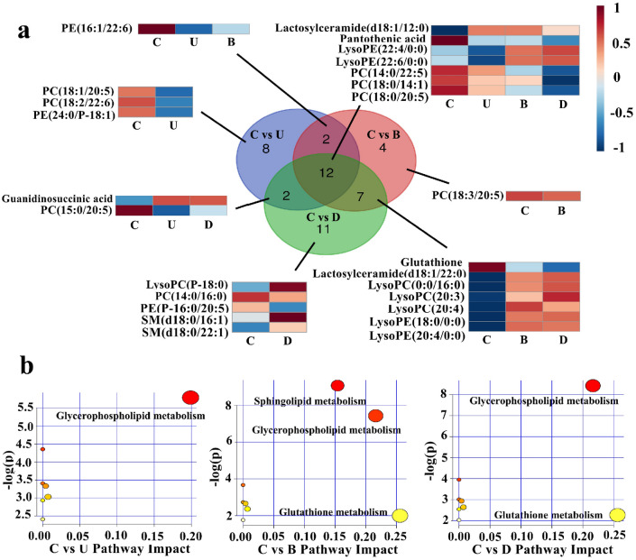
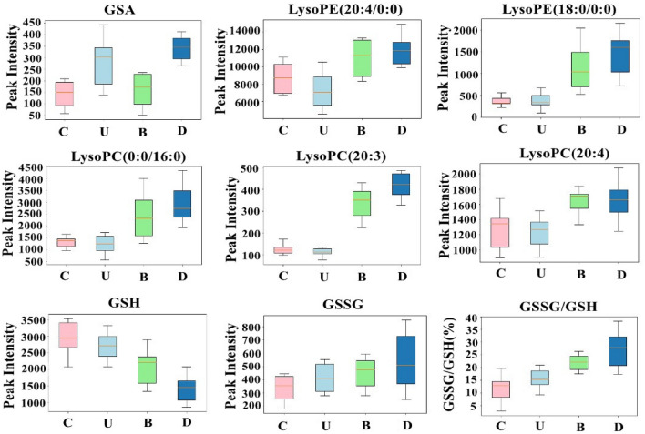
We have recently shown that both UVB and BaP can induce the production of ROS, apoptosis and even cancer. However, the differences in the metabolic profiles of skin damaged by UVB, BaP or UVB combined with BaP have not been studied. Therefore, we examined the metabolic changes in the human foreskin fibroblast injured by UVB or BaP or the combination of the two, using ultra performance liquid chromatography (UPLC) coupled with quadrupole time-of-flight mass spectrometry (qTOF-MS). 24 metabolites were altered in the UVB damage group, 25 in the BaP damage group, and 33 in the UVB combined with BaP group. These alterations indicated that the metabolic mechanisms of HFF-1 cells treated with UVB or BaP are related to multiple main metabolites including glycerophosphocholine (PC), lactosylceramide (LacCer), guanidinosuccinic acid (GSA), glutathione(GSH), and lysophosphatidylcholine (LysoPC) and the main mechanisms involved glycerophospholipid and glutathione metabolism. Thus, our report provided useful insight into the underlying mechanisms of UVB and BaP damage to skin cells.
Conflict of interest statement
The authors declare no competing interests.
Figures
Similar articles
-
Wavelength dependent responses of primary human keratinocytes to combined treatment with benzo[a]pyrene and UV light.Mutagenesis. 2005 Jul;20(4):305-10. doi: 10.1093/mutage/gei042. Epub 2005 Jun 14. Mutagenesis. 2005. PMID: 15956040
-
p53 gene mutations in SKH-1 mouse tumors differentially induced by UVB and combined subcarcinogenic benzo[a]pyrene and UVA.Photochem Photobiol. 2008 Mar-Apr;84(2):444-9. doi: 10.1111/j.1751-1097.2007.00282.x. Epub 2008 Jan 29. Photochem Photobiol. 2008. PMID: 18248503
-
Enzyme-modified Panax ginseng inhibits UVB-induced skin aging through the regulation of procollagen type I and MMP-1 expression.Food Funct. 2014 Feb;5(2):265-74. doi: 10.1039/c3fo60418g. Food Funct. 2014. PMID: 24281186
-
Mechanism of UVB-induced wrinkling of the skin: paracrine cytokine linkage between keratinocytes and fibroblasts leading to the stimulation of elastase.J Investig Dermatol Symp Proc. 2009 Aug;14(1):36-43. doi: 10.1038/jidsymp.2009.11. J Investig Dermatol Symp Proc. 2009. PMID: 19675551 Review.
-
Synergistic damage by UVA radiation and pollutants.Toxicol Ind Health. 2009 May-Jun;25(4-5):219-24. doi: 10.1177/0748233709106067. Toxicol Ind Health. 2009. PMID: 19651790 Review.
Cited by
-
Impact of Skin Exposure to Benzo[a]pyrene in Rat Model: Insights into Epidermal Cell Function and Draining Lymph Node Cell Response.Int J Mol Sci. 2024 Aug 8;25(16):8631. doi: 10.3390/ijms25168631. Int J Mol Sci. 2024. PMID: 39201318 Free PMC article.
-
Glutaminolysis impairment and immunometabolic dysregulation in U937 cells: Key mechanisms in occupational and environmental skin exposure to UV and benzo[a]pyrene.Arch Toxicol. 2025 Aug 25. doi: 10.1007/s00204-025-04155-4. Online ahead of print. Arch Toxicol. 2025. PMID: 40855183
-
Linoleic Acid Induces Metabolic Reprogramming and Inhibits Oxidative and Inflammatory Effects in Keratinocytes Exposed to UVB Radiation.Int J Mol Sci. 2024 Sep 26;25(19):10385. doi: 10.3390/ijms251910385. Int J Mol Sci. 2024. PMID: 39408715 Free PMC article.
-
UVA-induced metabolic changes in non-malignant skin cells and the potential role of pyruvate as antioxidant.Photochem Photobiol Sci. 2023 Aug;22(8):1889-1899. doi: 10.1007/s43630-023-00419-z. Epub 2023 May 17. Photochem Photobiol Sci. 2023. PMID: 37193818
-
Current Knowledge in Skin Metabolomics: Updates from Literature Review.Int J Mol Sci. 2022 Aug 7;23(15):8776. doi: 10.3390/ijms23158776. Int J Mol Sci. 2022. PMID: 35955911 Free PMC article. Review.
References
-
- Silveira JEPS, Pedroso DMM. UV light and skin aging. Rev. Environ. Health. 2014;29(3):243–254. - PubMed
-
- Fu PP, Xia Q, Xin S, et al. Phototoxicity and environmental transformation of polycyclic aromatic hydrocarbons (PAHs)—light-induced reactive oxygen species, lipid peroxidation, and dna damage. J Environ Sci Health C Environ Carcinog Ecotoxicol Rev. 2012;30(1):1–41. doi: 10.1080/10590501.2012.653887. - DOI - PubMed
-
- Rhodes, L. E. IARC Handbook of Cancer Prevention Sunscreens, vol. 5.: Harri Vainio and Franca Bianchini (Eds.); ISBN 92-832-3005-1. Surgical Oncology, 2001,10(1):72.
Publication types
MeSH terms
Substances
LinkOut - more resources
Full Text Sources
Other Literature Sources
Medical
