. 2021 May 27;11(1):11146.
doi: 10.1038/s41598-021-89983-x.

DHHC21 deficiency attenuates renal dysfunction during septic injury

Affiliations

DHHC21 deficiency attenuates renal dysfunction during septic injury

Xiaoyuan Yang et al. Sci Rep. .

Abstract

Renal dysfunction is one of the most common complications of septic injury. One critical contributor to septic injury-induced renal dysfunction is renal vascular dysfunction. Protein palmitoylation serves as a novel regulator of vascular function. Here, we examined whether palmitoyl acyltransferase (PAT)-DHHC21 contributes to septic injury-induced renal dysfunction through regulating renal hemodynamics. Multispectral optoacoustic imaging showed that cecal ligation and puncture (CLP)-induced septic injury caused impaired renal excretion, which was improved in DHHC21 functional deficient (Zdhhc21dep/dep) mice. DHHC21 deficiency attenuated CLP-induced renal pathology, characterized by tissue structural damage and circulating injury markers. Importantly, DHHC21 loss-of-function led to better-preserved renal perfusion and oxygen saturation after CLP. The CLP-caused reduction in renal blood flow was also ameliorated in Zdhhc21dep/dep mice. Next, CLP promoted the palmitoylation of vascular α1-adrenergic receptor (α1AR) and the activation of its downstream effector ERK, which were blunted in Zdhhc21dep/dep mice. Vasoreactivity analysis revealed that renal arteries from Zdhhc21dep/dep mice displayed reduced constriction response to α1AR agonist phenylephrine compared to those from wild-type mice. Consistently, inhibiting PATs with 2-bromopalmitate caused a blunted vasoconstriction response to phenylephrine in small arteries isolated from human kidneys. Therefore, DHHC21 contributes to impaired renal perfusion and function during septic injury via promoting α1AR palmitoylation-associated vasoconstriction.


Conflict of interest statement

The authors declare no competing interests.

Figures
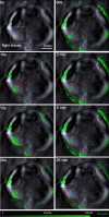
Figure 1
Figure 1
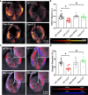
Representative MSOT images showing the clearance of IRDye800CW from mouse kidneys. IRDye800CW (60 nM dissolved in 0.9% saline) was injected intravenously into WT mice 24 h after sham operation. The clearance/movement of IRDye800CW (green) in the cross section of the right kidney (white square) was recorded. ROIs in cortex and medulla/pelvis region of the kidney are delineated in red and blue, respectively. Representative images of 28 mice (n = 7 in each group). The green bar represents the scale for the mean pixel intensity of the IRDye800CW signal.
Figure 2
Figure 2
Septic injury-induced delay in renal clearance of IRDye800CW is significantly attenuated in Zdhhc21dep/dep mice. (a) Representative IRDye800CW clearance kinetics of the cortex (red) and medulla/pelvis region (blue). Tmax-delay is the interval between the peaks of pixel intensity in cortex and medulla/pelvis region. (b) DHHC21 functional deficiency attenuates septic injury-induced increase in Tmax-delay. Results represent mean ± SEM. n = 7, *p ≤ 0.05 versus WT + Sham, #p ≤ 0.05 versus WT + CLP.
Figure 3
Figure 3
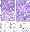
Zdhhc21dep/dep mice are resistant to septic injury-induced kidney damage. (a) Representative images of Periodic Acid-Schiff-stained renal tissue collected 24 h after septic injury induction. Pathological alterations are indicated by arrows with different colors: brush border loss (blue), tubular cell detachment (red), neutrophil infiltration (black). Images are representative of 5 mice. (b) Mice with DHHC21 loss-of-function display a reduced renal injury score than WT mice after septic injury. n = 5 independent experiments; 5 views per animal were imaged and analyzed. (c) The level of circulating creatinine. Mouse plasma was collected 24 h after CLP-induced septic injury. n = 12. (d) The level of blood urea nitrogen 24 h after septic injury. n = 11. Results represent mean ± SEM. *p ≤ 0.05 versus WT + Sham, #p ≤ 0.05 versus WT + CLP.
Figure 4
Figure 4
DHHC21 functional deficiency prevents the reduction of renal perfusion and renal oxygen saturation after septic injury. (a) Representative MSOT images showing the intensity of total hemoglobin in kidneys. MSOT images are pseudo-colored with hot-color scale. (b) Higher signal intensity of total hemoglobin is observed in kidneys of Zdhhc21dep/dep mice than WT ones after septic injury. n = 9. (c) Reconstructed MSOT images indicate the distribution of oxygenated hemoglobin (red) and deoxygenated hemoglobin (blue) in kidney cross sections 24 h after CLP induction. (d) Zdhhc21dep/dep mice display higher level of renal SO2 compared to WT mice after septic injury. ROIs are drawn around right kidneys. n = 8. Results represent mean ± SEM. *p ≤ 0.05 versus WT + Sham, #p ≤ 0.05 versus WT + CLP.
Figure 5
Figure 5
DHHC21 loss-of-function attenuates septic injury-induced reduction in renal blood flow. (a,b) Representative recordings of RBF (a) and MAP (b) in different groups 24 h after septic injury. Images are representative of 8 mice. (c) The reduced RBF in WT mice upon septic injury is rescued in Zdhhc21dep/dep mice. n = 8 (d) No significant difference in MAP is observed between WT and Zdhhc21dep/dep mice upon septic injury. n = 8. Results represent mean ± SEM. *p ≤ 0.05 versus WT + Sham, #p ≤ 0.05 versus WT + CLP.
Figure 6
Figure 6
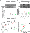
DHHC21 catalyzes α1AR palmitoylation and contributes to α1AR-mediated signaling pathway activation and vasoconstriction. (a) The level of palmitoylated α1AR in renal arteries. Palmitoylated proteins were isolated by RAC in the presence of hydroxylamine and then analyzed via immunoblotting for α1AR. Blots are representative of 8 independent experiments; renal arteries from 12 mice were pooled for each independent immunoblot analysis. Band intensity is quantified and normalized to control. Full-length blots are presented in Supplementary Fig. S5a. (b) Septic injury-induced ERK activation is inhibited in Zdhhc21dep/dep mice. The band intensity of phosphorylated ERK is normalized to that of total ERK. n = 9. *p ≤ 0.05 versus WT + Sham, #p ≤ 0.05 versus WT + CLP. Full-length blots are presented in Supplementary Fig. S5b. (c) Representative myograph recordings of WT and Zdhhc21dep/dep renal arteries. KPSS represents 60 mM potassium physiological saline solution; W represents wash. (d) Myograph results show that renal arteries of Zdhhc21dep/dep mice exhibit reduced tension compared to those of WT mice upon phenylephrine challenge. Results represent mean ± SEM. n = 9 mice. One artery per animal was used. *p ≤ 0.05 versus WT.
Figure 7
Figure 7
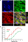
DHHC21 co-localizes with α1AR and palmitoylation regulates the constriction of small arteries isolated from human kidneys. (a) Co-localization of DHHC21 and α1AR in the cross-section of human renal arteries. Representative images of renal arteries isolated from 3 donors. (b) Inhibition of PATs with 2-BP results in impaired α1AR-mediated vasoconstriction in small arteries isolated from human kidneys. Vehicle control or 2-BP (100 μM) was given 1 h prior to phenylephrine challenge. Small arteries are from 3 different donors. Results represent mean ± SEM. *p ≤ 0.05.

Similar articles

Cited by

References

    1. Case J, Khan S, Khalid R, Khan A. Epidemiology of acute kidney injury in the intensive care unit. Crit. Care Res. Pract. 2013;2013:479730. doi: 10.1155/2013/479730. - DOI - PMC - PubMed
    1. Godin M, Murray P, Mehta RL. Clinical approach to the patient with AKI and sepsis. Semin. Nephrol. 2015;35:12–22. doi: 10.1016/j.semnephrol.2015.01.003. - DOI - PMC - PubMed
    1. Doi K. Role of kidney injury in sepsis. J. Intensive Care. 2016;4:17. doi: 10.1186/s40560-016-0146-3. - DOI - PMC - PubMed
    1. Zarjou A, Agarwal A. Sepsis and acute kidney injury. J. Am. Soc. Nephrol. 2011;22:999–1006. doi: 10.1681/ASN.2010050484. - DOI - PubMed
    1. Ronco C, Kellum JA, Bellomo R, House AA. Potential interventions in sepsis-related acute kidney injury. Clin. J. Am. Soc. Nephrol. 2008;3:531–544. doi: 10.2215/CJN.03830907. - DOI - PMC - PubMed

Publication types

Substances