. 2021 May 27;11(1):11222.
doi: 10.1038/s41598-021-90809-z.

Interactions between the flavescence dorée phytoplasma and its insect vector indicate lectin-type adhesion mediated by the adhesin VmpA

Affiliations

Interactions between the flavescence dorée phytoplasma and its insect vector indicate lectin-type adhesion mediated by the adhesin VmpA

Nathalie Arricau-Bouvery et al. Sci Rep. .

Abstract

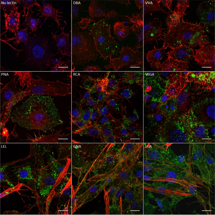
The flavescence dorée phytoplasma undergoes a propagative cycle in its insect vectors by first interacting with the insect cell surfaces, primarily in the midgut lumen and subsequently in the salivary glands. Adhesion of flavescence dorée phytoplasma to insect cells is mediated by the adhesin VmpA. We hypothesize that VmpA may have lectin-like activity, similar to several adhesins of bacteria that invade the insect gut. We first demonstrated that the luminal surface of the midgut and the basal surface of the salivary gland cells of the natural vector Scaphoideus titanus and those of the experimental vector Euscelidius variegatus were differentially glycosylated. Using ELISA, inhibition and competitive adhesion assays, and protein overlay assays in the Euva-6 insect cell line, we showed that the protein VmpA binds insect proteins in a lectin-like manner. In conclusion, the results of this study indicate that N-acetylglucosamine and mannose present on the surfaces of the midgut and salivary glands serve as recognition sites for the phytoplasma adhesin VmpA.


Conflict of interest statement

The authors declare no competing interests.

Figures

Figure 1
Figure 1
Observation of ingested fluorescent lectins binding midgut carbohydrates of E. variegatus by laser scanning microscopy. Cell nuclei were stained with DAPI (blue), and actin filaments were stained with Alexa 568-phalloidin (red). The FITC-lectins were colored in green. The fluorescent lectins used to stain the cells are indicated in each picture except for the control, where fluorescent lectin was omitted. amg anterior midgut, mmg middle midgut, pmg posterior midgut, fc filter chamber, mt Malpighian tubules. Arrowheads in panels DBA and PNA indicate the presence of fluorescence. Scale bar 100 µm.
Figure 2
Figure 2
Observation of fluorescent lectins binding salivary gland cell carbohydrates of E. variegatus by laser scanning microscopy. Cell nuclei were stained with DAPI (blue), and actin filaments were stained with Alexa 568-phalloidin (red). The FITC-lectins were colored in green. The fluorescent lectins used to stain the cells are indicated in each picture except for the control, where fluorescent lectin was omitted. The arrowhead indicates one of the two cell types that possess glycoconjugates bound by the WGA lectin, and the arrows indicate the other cell type. Scale bar 100 µm.
Figure 3
Figure 3
Observation by laser scanning microscopy of fluorescent lectins binding salivary gland carbohydrates of FDP-infected E. variegatus. Cell nuclei were stained with DAPI (blue), actin filaments with Alexa 568-phalloidin (red), phytoplasmas with anti-VmpA antibodies and Alexa 633-secondary antibodies (cyan), and FITC-lectins are colored green. (a) Overlay pictures of infected salivary gland cells stained with the lectin indicated in each picture. Arrows indicate the FDP-infected cells, and arrowheads indicate the cells recognized by the lectin used. (b) View of the infected salivary gland surfaces stained with the lectin indicated in each picture. Scale bar 100 µm.
Figure 4
Figure 4
Magnification of salivary glands of FDP-infected E. variegatus stained with fluorescent LEL, GNA and LCA lectins by laser scanning microscopy with an Airyscan detector. FDPs were stained with anti-VmpA antibodies and Alexa 633-secondary antibodies and (cyan), cell nuclei with DAPI (blue), actin filaments with Alexa 568-phalloidin (red) and glycoconjugates with FITC-lectins LEL, GNA and LCA (green). (a) View of the infected salivary gland cell surface. (b) View of the inside of infected salivary gland cells. Arrows indicate structures resembling apical membranes described by Koinuma et al. around which phytoplasmas are grouped. Arrowheads indicate phytoplasmas that crossed the apical plasma membrane. Scale bar 10 µm.
Figure 5
Figure 5
Observation of fluorescent lectins binding Euva-6 cell carbohydrates by laser scanning microscopy with an Airyscan detector. Cell nuclei were stained with DAPI (blue), actin filaments with Alexa 568-phalloidin (red) and glycoconjugates with FITC-lectins (green). The fluorescent lectins used to stain the cells are indicated in each picture except for the control, where fluorescent lectin was omitted. Scale bar 10 µm.
Figure 6
Figure 6
Interaction of VmpA with Euva-6 cells in the presence of sugar and lectins. (a) Adhesion of VmpA-His6 proteins to Euva-6 cells measured by ELISA in the presence of sugars. The recombinant proteins were first incubated with different concentrations of sugars indicated below the graphs before being added to Euva-6 cells. Boxplots with different letters are significantly different under the ANOVA test of the R commander package of R software version 4.0.3 (R: A language and Environment for statistical computing, R Core Team, R Foundation for Statistical Computing, Vienna, Austria, 2020, https://www.R-project.org). The normal distribution of the value was previously verified using the Shapiro–Wilk normality test of R software (R commander package). (b) Adhesion of VmpA-His6-coated beads to Euva-6 cells in competition with lectins. Euva-6 cells were preincubated with the lectins indicated above the graphs before being incubated with fluorescent VmpA-His6-coated beads. 0 indicates that no lectin was incubated with insect cells. The P values according to the Kruskal–Wallis rank sum test of the software R (R commander package) are indicated above the boxplots.
Figure 7
Figure 7
Interaction of VmpA-His6 with insect proteins. The results are presented from different blots (full length) framed by black lines. (a) Euva-6 proteins from culture passage 7 transferred to nitrocellulose membranes were incubated with recombinant VmpA-His6 alone (lane 0) or in the presence of 0.1 to 1 M Gal, Glc, GlcNAc or Man. The presence of VmpA-His6 interacting with insect cells was detected using anti-VmpA antibodies. (b) Euva-6 proteins from culture passage 32 transferred to nitrocellulose membranes were incubated with recombinant VmpA-His6 or the lectins LEL and GNA.

Similar articles

Cited by

References

    1. Lee I-M, Davis RE, Gundersen-Rindal DE. Phytoplasma: Phytopathogenic mollicutes. Annu. Rev. Microbiol. 2000;54:221–255. doi: 10.1146/annurev.micro.54.1.221. - DOI - PubMed
    1. Gasparich GE. Spiroplasmas and phytoplasmas: Microbes associated with plant hosts. Biol. J. Int. Assoc. Biol. Stand. 2010;38:193–203. - PubMed
    1. Namba S. Molecular and biological properties of phytoplasmas. Proc. Jpn. Acad. B. 2019;95:401–418. doi: 10.2183/pjab.95.028. - DOI - PMC - PubMed
    1. Sugio A, et al. Diverse targets of phytoplasma effectors: From plant development to defense against insects. Annu. Rev. Phytopathol. 2011;49:175–195. doi: 10.1146/annurev-phyto-072910-095323. - DOI - PubMed
    1. Caudwell A. Epidemiology and characterization of flavescence-doree (fd) and other grapevine yellows. Agronomie. 1990;10:655–663. doi: 10.1051/agro:19900806. - DOI

Publication types

LinkOut - more resources