. 2018 Mar 21;28(12):1701183.
doi: 10.1002/adfm.201701183. Epub 2017 Sep 4.

Engineered Axonal Tracts as "Living Electrodes" for Synaptic-Based Modulation of Neural Circuitry

Affiliations

Engineered Axonal Tracts as "Living Electrodes" for Synaptic-Based Modulation of Neural Circuitry

Mijail D Serruya et al. Adv Funct Mater. .

Abstract

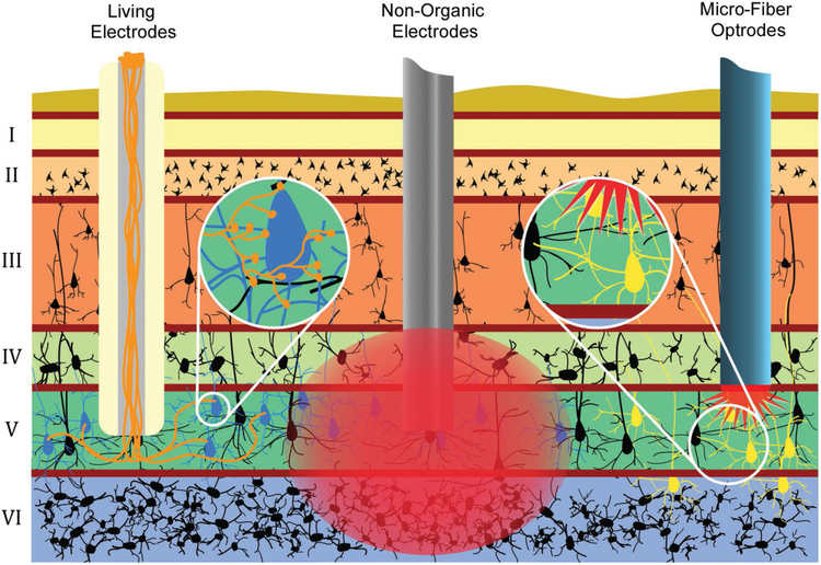
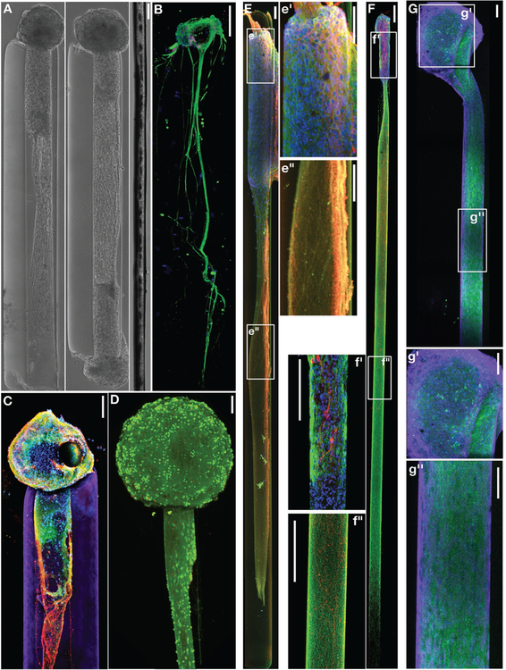
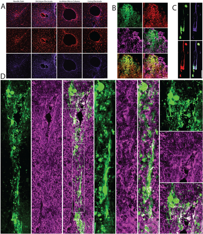
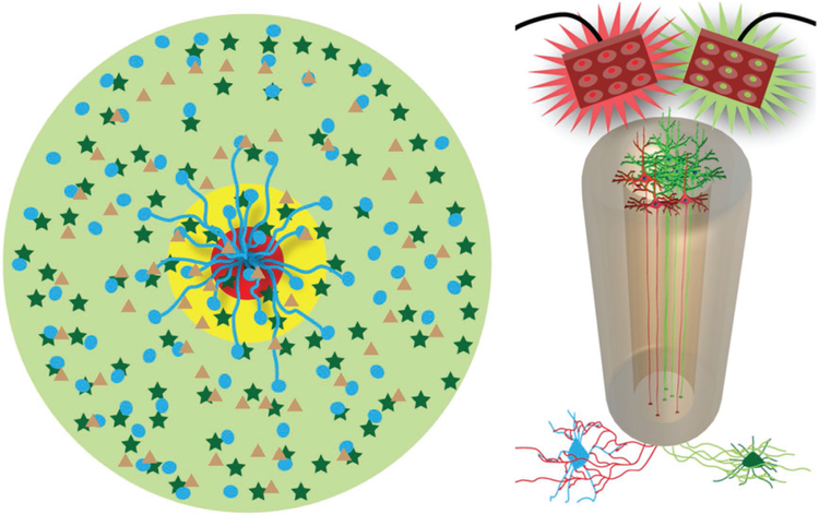
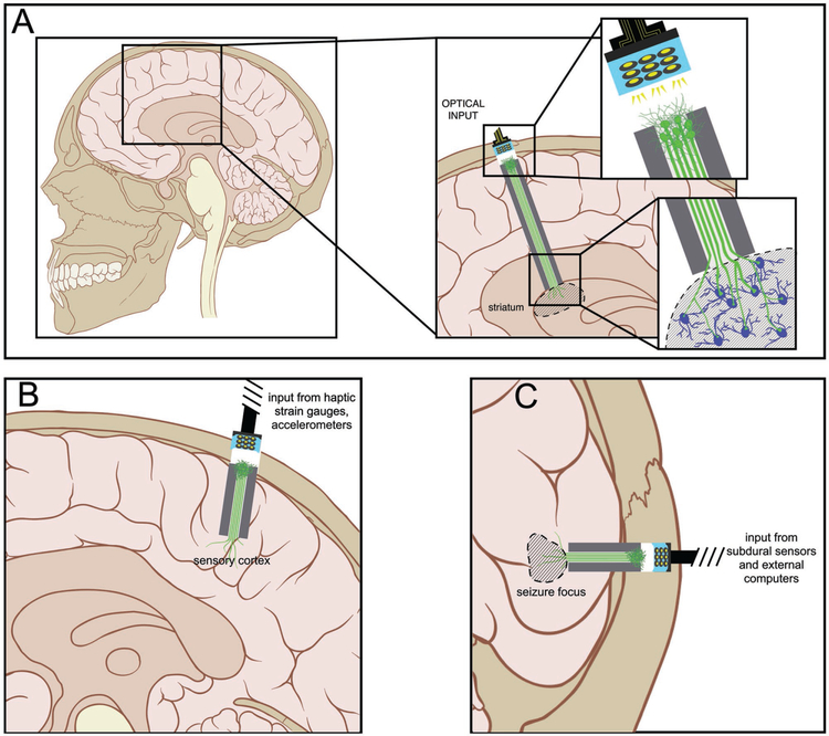
Brain-computer interface and neuromodulation strategies relying on penetrating non-organic electrodes/optrodes are limited by an inflammatory foreign body response that ultimately diminishes performance. A novel "biohybrid" strategy is advanced, whereby living neurons, biomaterials, and microelectrode/optical technology are used together to provide a biologically-based vehicle to probe and modulate nervous-system activity. Microtissue engineering techniques are employed to create axon-based "living electrodes", which are columnar microstructures comprised of neuronal population(s) projecting long axonal tracts within the lumen of a hydrogel designed to chaperone delivery into the brain. Upon microinjection, the axonal segment penetrates to prescribed depth for synaptic integration with local host neurons, with the perikaryal segment remaining externalized below conforming electrical-optical arrays. In this paradigm, only the biological component ultimately remains in the brain, potentially attenuating a chronic foreign-body response. Axon-based living electrodes are constructed using multiple neuronal subtypes, each with differential capacity to stimulate, inhibit, and/or modulate neural circuitry based on specificity uniquely afforded by synaptic integration, yet ultimately computer controlled by optical/electrical components on the brain surface. Current efforts are assessing the efficacy of this biohybrid interface for targeted, synaptic-based neuromodulation, and the specificity, spatial density and long-term fidelity versus conventional microelectronic or optical substrates alone.

Keywords: biologically-mediated neuromodulation; brain–computer interfaces; living scaffolds; microtissue engineering; tissue engineering.


Conflict of interest statement

Conflict of Interest The authors declare no conflict of interest.

Figures

Figure 1.
Figure 1.
Advantages of Axon-Based “Living Electrodes” for Neuromodulation: Mechanisms and specificity of neuronal stimulation for “living electrodes” (left) versus conventional electrodes (center) and optrodes (right). Living electrodes provide engineered axonal tracts, fully differentiated neurons, and a controlled 3D cytoarchitecture, potentially improving survival versus delivery of cell suspensions. Construct neurons may be transfected to express opsins in vitro (days prior to implant), thereby avoiding the injection of virus directly into the host while constraining the spatial extent of transfected cells. Living electrodes could offer high specificity, as the constructs can be designed to synapse with specific neuronal subtypes in a given anatomical region (as shown by living electrode axons synapsing with only blue neurons, not black) as opposed to conventional electrodes that inherently stimulate or record from a relatively large 3D volume around the electrode (as shown by large red area of stimulation affecting many layers and neurons). While optrodes can achieve a high level of specificity, the in vivo delivery of opsins generally relies on injection of virus that may diffuse and affect non-target regions (spread of optogenetic transduction is illustrated by yellow neurons in multiple layers). Also, optical methods may have a limited extent due to tissue absorption of light. Finally, living electrodes provide a soft pathway to route signals to/from deep brain structures compared to rigid materials used in electrodes/optrodes, thus potentially minimizing signal loss due to mechanical mismatch/micromotion and glial scarring.
Figure 2.
Figure 2.
Neuronal-Axonal Living Electrodes: (A) Phase contrast images of unidirectional (left) and bidirectional (middle) “living electrodes” built using cerebral cortical neurons, each at 5 days in vitro (DIV), next to a single human hair (right). (B) Confocal reconstruction of a living electrode built using dorsal root ganglia neurons showing unidirectional axonal tracts immunolabeled to denote neuronal somata (MAP-2; purple) and axons (tau; green), with nuclear counterstain (blue). (C) Confocal reconstruction of a unidirectional, cerebral cortical neuronal living electrode at 11 DIV, immunolabeled for axons (β-tubulin-III; red) and synapses (synapsin; green), with a nuclear counterstain (Hoechst; blue). The surrounding hydrogel micro-column is shown in purple. (D) Confocal reconstruction of a unidirectional cortical neuronal living electrode stained for viability at 10 DIV (green: live cells via calcein-AM; red: nuclei of dead cells via ethidium homodimer-1). Scale bars A-D: 100 μm. (E-G) Long-projecting unidirectional axon-based living electrodes for tailored neuromodulation. (E) Confocal reconstruction of an excitatory living electrode built using neurons derived from the cerebral cortex (predominantly glutamatergic), immunolabeled at 28 DIV for axons (β-tubulin-III; red) and neuronal somata/dendrites (MAP-2; green), with nuclear counterstain (Hoechst; blue). Insets of the aggregate (e’) and axonal (e”) regions are outlined and shown to the right. Scale bars: 100 μm. (F) Confocal reconstruction of a dopaminergic living electrode built using neurons isolated from the ventral mesencephalon (enriched in dopaminergic neurons), immunolabeled at 28 DIV for axons (β-tubulin-III; green) and tyrosine hydroxylase (dopaminergic neurons/axons; red), with nuclear counterstain (Hoechst; blue). Insets of the aggregate (f’) and axonal (f”) regions are outlined and shown to the left. Scale bars: 250 μm. (G) Confocal reconstruction of an inhibitory living electrode built using neurons isolated from the medial ganglionic eminence (source of GABAergic neurons), immunolabeled at 14 DIV for axons (β-tubulin-III; purple) and GABA (inhibitory neurons/axons; green), with nuclear counterstain (Hoechst; blue). Insets of the aggregate (g’) and axonal (g”) regions are outlined and shown below. Scale bars: 100 μm.
Figure 3.
Figure 3.
Neuronal Survival, Synaptic Integration, and Host Response Following Living Electrode Implantation In Vivo: (A) Host response to living electrodes versus conventional microelectrodes. Representative confocal micrographs at 1-month post-implant of brain sections orthogonal to a needle stab (negative control), a Michigan microelectrode (positive control), acellular hydrogel micro-column, or a living electrode (hydrogel micro-column encasing neurons + axonal tracts) immunolabeled for microglia/macrophages (IBA-1; red) and astrocytes (GFAP; purple). Peri-electrode host reactivity was reduced around living electrodes, even though current-generation living electrodes have a larger footprint than Michigan microelectrodes. (B-D) Confocal reconstructions showing survival and integration of living electrode neurons/axons at 1-week or 1-month post-implant. (B) Superficial (dorsal) living electrode neurons on the brain surface transduced to express GFP (on the synapsin promoter; green) and immunolabeled for the neuronal marker NeuN (red) and the synaptic marker synapsin (purple) with various dual- and tri-channel combinations. (C) Living electrode neurons and aligned axons (GFP+) within the lumen of the micro-column stained to identify neuronal somata and dendrites (MAP-2; red) and axons (β-tubulin-III; purple). (D) Neurons and neurites projecting in the cerebral cortex from the deep end of the living electrode, with callout boxes showing putative synapses (synapsin+ puncta; purple) between host and living electrode neurons/neurites (GFP+). Scale bars: 50 μm.
Figure 4.
Figure 4.
Mechanisms-of-Action for Axon-Based Living Electrodes: Synaptic Specificity, Biological Multiplexing, and Stability. “Living electrodes” may offer high specificity, as the constructs can be designed to synapse with specific neuronal subtypes, as demonstrated conceptually by living electrode axons synapsing with only circle neurons, not star neurons (left cartoon). This may be exploited in mixed neuron living electrodes where a subpopulation (blue cells) is excited with red light while another subpopulation (dark green cells) could be inhibited by green light (right cartoon). Multiplexing: one living electrode axon can (in theory) synapse with hundreds to thousands of host neurons – creating a significant amplification effect. We currently build living electrodes with 5000–50 000 neurons within a column less than twice the diameter of a human hair. Moreover, living electrodes may offer stability as synaptic integration offers permanence not possible with standard approaches while the biological nature of the constructs may mitigate the chronic foreign body response.
Figure 5.
Figure 5.
Potential Applications of Axon-Based Living Electrodes: Custom engineered living electrodes consisting of a phenotypically-controlled populations of neurons extending long axonal tracts through a biocompatible micro-column may be stereotactically transplanted to span various regions to treat particular disease processes. (A) Axons projecting from dopaminergic living electrodes may form synapses within local striatal architecture, and, due to in vitro functionalization with channelrhodopsins, may release dopamine upon optical stimulation of the perikaryal segment at the brain surface. This mimics the substantia nigra pars compacta input to the striatum in a manner that can be externally controlled. (B) Axons from glutamatergic living electrodes may preferentially synapse onto layer IV neurons within primary sensory cortex to convey illusory haptic feedback via surface optical stimulation to achieve closed-loop control of neuromotor prosthetics in patients with paralysis. (C) Axons from GABAergic living electrodes could be implanted to oppose seizure foci such that optical stimulation would cause net suppression of seizure activity in patients with lesional epilepsy.
Figure 6.
Figure 6.
Exploiting “Biological Multiplexing” in Living Electrodes. More sophisticated living electrodes may be developed to further exploit so-called biological multiplexing. By fabricating the constructs in vitro using microprinting and micropatterning techniques, specific synaptic architectures can be achieved to yield certain fine-grained signal manipulations linking the construct to the brain. (A) In the simplest form, “channel select” bundles of axons can transmit signals to select which other bundles transmit signals into the brain, and which are silenced. (B) Multiple channels that converge on to one final common output can likewise be toggled by the “channel select” in a biological instantiation that most resembles the kind of multiplexing used in telecommunications. (C) Likewise, a single input channel can be selected and diverted to one or more parallel outputs to “demultiplex” that signal.
Figure 7.
Figure 7.
Potential for Time-Division “Biological Multiplexing” in Living Electrodes. Future iterations of living electrodes may exploit delay lines emanating from a single “clock” circuit formed by a cluster of neurons linked by gap junctions (coupled damped oscillators) and micropatterned inhibitory and excitatory connections. Thus, multiple parallel input channels can be multiplexed serially with each clock cycle to a single target output neuron that in turn links to the brain. The rate of the clock (and hence the multiplexing sampling duration) can be altered by driving the clock circuit directly.

Similar articles

Cited by

References

    1. Struzyna LA, Wolf JA, Mietus CJ, Adewole DO, Chen HI, Smith DH, Cullen DK, Tissue Eng. Part A 2015, 21, 2744. - PMC - PubMed
    1. Struzyna LA, Harris JP, Katiyar KS, Isaac Chen H, Cullen DK, Neural Regen. Res 2015, 10, 679. - PMC - PubMed
    1. Duda JE, Struzyna LA, Browne KD, Chen HI, Wolf JA, Cullen DK, Proceedings of 21st International Congress of Parkinson’s Disease and Movement Disorders 2017.
    1. Struzyna LA, Adewole DO, Gordián-Vélez WJ, Grovola MR, Burrell JC, Katiyar KS, Petrov D, Harris JP, Cullen DK,J. Vis. Exp 2017, 123, e55609. - PMC - PubMed
    1. Harris JP, Struzyna LA, Murphy PL, Adewole DO, Kuo E, Cullen DK, J. Neural Eng 2016, 13, 16019. - PMC - PubMed

LinkOut - more resources