. 2021 May 11:12:652860.
doi: 10.3389/fphar.2021.652860. eCollection 2021.

Zerumbone Suppresses the LPS-Induced Inflammatory Response and Represses Activation of the NLRP3 Inflammasome in Macrophages

Affiliations

Zerumbone Suppresses the LPS-Induced Inflammatory Response and Represses Activation of the NLRP3 Inflammasome in Macrophages

Chia-Cheng Su et al. Front Pharmacol. .

Abstract

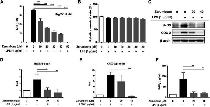
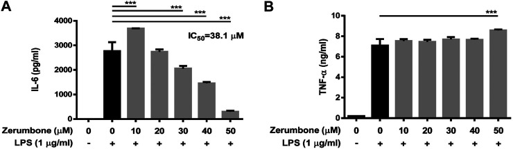
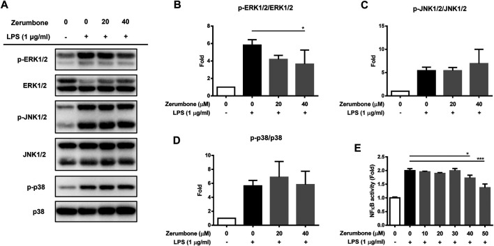
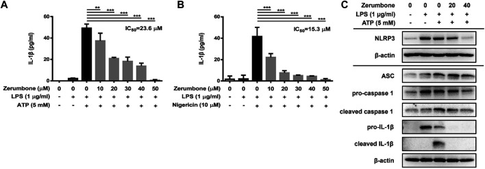
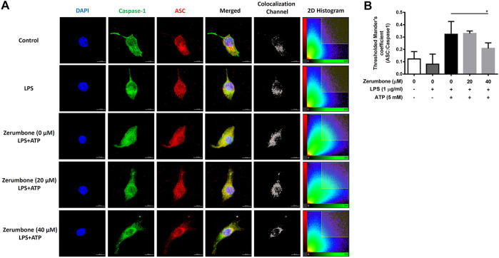
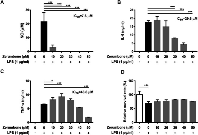
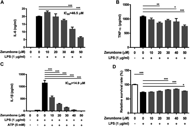
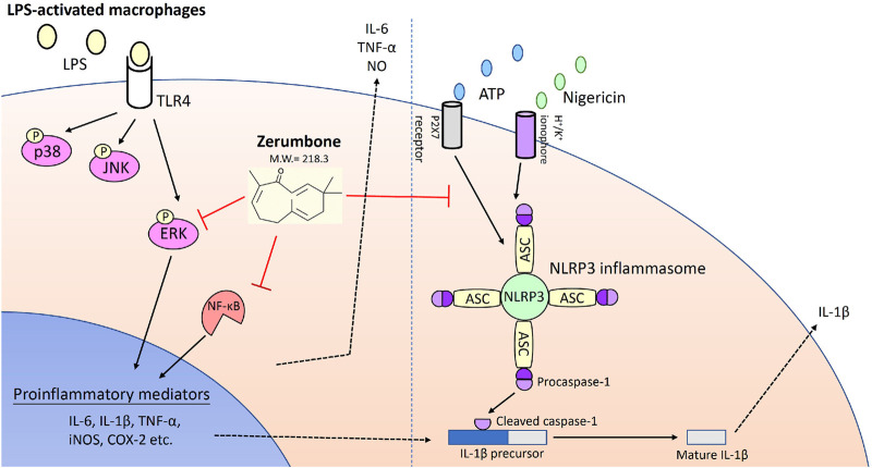
Zerumbone is a natural product isolated from the pinecone or shampoo ginger, Zingiber zerumbet (L.) Smith, which has a wide range of pharmacological activities, including anti-inflammatory effects. However, the effects of zerumbone on activation of the NLRP3 inflammasome in macrophages have not been examined. This study aimed to examine the effects of zerumbone on LPS-induced inflammatory responses and NLRP3 inflammasome activation using murine J774A.1 cells, murine peritoneal macrophages, and murine bone marrow-derived macrophages. Cells were treated with zerumbone following LPS or LPS/ATP treatment. Production of nitric oxide (NO) was measured by Griess reagent assay. The levels of IL-6, TNF-α, and IL-1β secretion were analyzed by ELISA. Western blotting analysis was performed to determine the expression of inducible NO synthase (iNOS), COX-2, MAPKs, and NLRP3 inflammasome-associated proteins. The activity of NF-κB was determined by a promoter reporter assay. The assembly of NLRP3 was examined by immunofluorescence staining and observed by confocal laser microscopy. Our experimental results indicated that zerumbone inhibited the production of NO, PGE2 and IL-6, suppressed the expression of iNOS and COX-2, repressed the phosphorylation of ERK, and decreased the activity of NF-κB in LPS-activated J774A.1 cells. In addition, zerumbone suppressed the production of IL-1β and inhibited the activity of NLRP3 inflammasome in LPS/ATP- and LPS/nigericin-activated J774A.1 cells. On the other hand, we also found that zerumbone repressed the production of NO and proinflammatory cytokines in LPS-activated murine peritoneal macrophages and bone marrow-derived macrophages. In conclusion, our experimental results demonstrate that zerumbone effectively attenuates the LPS-induced inflammatory response in macrophages both in vitro and ex vivo by suppressing the activation of the ERK-MAPK and NF-κB signaling pathways as well as blocking the activation of the NLRP3 inflammasome. These results imply that zerumbone may be beneficial for treating sepsis and inflammasome-related diseases.

Keywords: MAPKs; NLRP3 inflammasome; inflammation; macrophage; zerumbone.


Conflict of interest statement

The authors declare that the research was conducted in the absence of any commercial or financial relationships that could be construed as a potential conflict of interest.

Figures

FIGURE 1
FIGURE 1
The effects of zerumbone on the NO production, cell viability, and iNOS and COX-2 expressions in LPS-activated J774A.1 cells. Cells were pre-treated with various doses of zerumbone for 1 h following treatment of LPS (1 μg/ml) for 24 h. (A) The cell culture supernatant was collected and the production of NO was determined by Griess reagent assay. (B) Cell viability was analyzed by MTT assay. (C) Cells were harvested and lyzed by lysis buffer, and the expressions of iNOS and COX-2 were determined using Western blot. β-actin was regarded as a loading control. The representative Western blot results were obtained in three separate experiments. (D–E) The intensities of bands were quantified from three separate experiments and normalized to untreated samples. (F) Cell supernatants were collected and the levels of PGE2 were measured by ELISA. Data were represented as means ± SD. Statistical significance between the zerumbone-treated groups vs. the LPS-only group is represented as follows: *p < 0.05, **p < 0.01 and ***p < 0.001.
FIGURE 2
FIGURE 2
The effects of zerumbone on the production of IL-6 and TNF-α in LPS-activated J774A.1 cells. Cells were pre-treated with various doses of zerumbone for 1 h following treatment of LPS (1 μg/ml) for 24 h. Cell supernatants were collected and the levels of (A) IL-6 and (B) TNF-α were analyzed using ELISA. Data were collected in triplicate and represented as means ± SD. Statistical significance between the zerumbone-treated groups vs. the LPS-only group is represented as follows: ***p < 0.001.
FIGURE 3
FIGURE 3
The effects of zerumbone on the activation of MAPK and NF-κB in LPS-activated J774A.1 cells. Cells were pre-treated with various doses of zerumbone for 1 h following treatment of LPS (1 μg/ml) for 2 h. (A) The expressions of MAPK-associated proteins were determined by Western blot. Phospho-specific signals were then normalized against the total level of the target protein, using that protein as its own internal loading control for maximum accuracy. The representative Western blot results were obtained in three separate experiments. (B–D) The intensities of bands were quantified from three separate experiments and normalized to untreated samples. Data were resented as means ± SD. (D) J-blue cells, a stable NF-κB reporter cell line generated by transfected J774A.1 cells with NF-κB SEAP report plasmids, were pre-treated with various doses of zerumbone for 1 h following treatment of LPS (1 μg/ml) for 6 h. The activation of NF-κB was examined by SEAP activity. Data were represented as means ± SD. Statistical significance between the zerumbone-treated groups vs. the LPS-only group is represented as follows: *p < 0.05, **p < 0.01 and ***p < 0.001.
FIGURE 4
FIGURE 4
The effects of zerumbone on the production of IL-1β and the activity of NLRP3 inflammasome in LPS/ATP- and LPS/nigericin-activated J774A.1 cells. Cells were pre-treated with various doses of zerumbone for 1 h and then treated with LPS (1 μg/ml) for 5 h following treatment of ATP (5 mM) or nigericin (10 μΜ) for 30 min. (A, B) Cell culture supernatants were collected and the levels of IL-1β were determined by ELISA. Data were represented as means ± SD. Statistical significance between the zerumbone-treated groups vs. the LPS/ATP or LPS/nigericin groups is represented as follows: *p < 0.05, **p < 0.01 and ***p < 0.001. (C) The expression levels of inflammasome-associated proteins were analyzed by Western blot. β-actin was regarded as a loading control. The representative Western blot results were obtained in three separate experiments.
FIGURE 5
FIGURE 5
The effects of zerumbone on the colocalization of NLRP3 inflammasome components (ASC and caspase-1) in LPS/ATP-activated J774 A.1 cells. Cells were pre-treated with various doses of zerumbone for 1 h and then treated with LPS (1 μg/ml) for 5 h following treatment of ATP (5 mM) for 30 min. (A) Cells were stained with caspase-1 (green), ASC (red), and DAPI (blue) and then analyzed using confocal microscopy and Imaris software for confocal image 3D reconstruction (B) ASC speck formation was analyzed by the threshold of 2D histogram in panel A to determine the colocalization of caspase-1 and ASC signals using Mander’s coefficient. Data were represented as means ± SD. Statistical significance between the zerumbone-treated groups vs. the LPS/ATP group is represented as follows: *p < 0.05.
FIGURE 6
FIGURE 6
The effects of zerumbone on the NO, IL-6, and TNF-α productions and cell viability in LPS-activated murine peritoneal macrophages. Cells were pre-treated with various doses of zerumbone for 1 h following treatment of LPS (1 μg/ml) for 24 h. (A) The production of NO was determined by Griess reagent assay. The expression levels of (B) IL-6 and (C) TNF-α were measured using ELISA. Statistical significance between the zerumbone-treated groups vs. the LPS-only group is represented as follows: *p < 0.05 and ***p < 0.001. (D) Cell viability was analyzed by MTT assay. Data were represented as means ± SD. Statistical significance is represented as follows: ***p < 0.001, compared with the LPS-only group.
FIGURE 7
FIGURE 7
The effects of zerumbone on the cell viability and IL-6, TNF-α, and IL-1β productions in LPS- or LPS/ATP-activated murine bone marrow-derived macrophages. Cells were pre-treated with various doses of zerumbone for 1 h following treatment of LPS (1 μg/ml) for 24 h. The expression levels of (A) IL-6 and (B) TNF-α were measured using ELISA. Statistical significance between the zerumbone-treated groups vs. the LPS-only group is represented as follows: *p < 0.05, **p < 0.01 and ***p < 0.001. (C) For the IL-1β secretion, cells were pre-treated with various doses of zerumbone for 1 h and then treated with LPS (1 μg/ml) for 5 h following treatment of ATP (5 mM) for 30 min. Cell culture supernatants were collected and the levels of IL-1β were determined by ELISA. Data were represented as means ± SD. Statistical significance between the zerumbone-treated groups vs. the LPS-only group is represented as follows: ***p < 0.001. (D) Cell viability was analyzed by MTT assay. Statistical significance between the zerumbone-treated groups vs. the LPS-only group is represented as follows: *p < 0.05 and ***p < 0.001.
FIGURE 8
FIGURE 8
Zerumbone suppresses the LPS-induced inflammatory response and represses activation of the NLRP3 inflammasome in macrophages. Zerumbone effectively attenuates LPS-induced inflammatory responses in macrophages both in vitro and ex vivo by suppressing activation of the ERK-MAPK and NF-κB signaling pathways, inhibiting LPS-induced inflammatory mediators, and blocking the activation of the NLRP3 inflammasome.

Similar articles

Cited by

References

    1. Al-Amin M., Sultana G. N. N., Hossain C. F. (2012). Antiulcer Principle from Zingiber Montanum. J. Ethnopharmacology 141, 57–60. 10.1016/j.jep.2012.01.046 - DOI - PubMed
    1. Banerjee A., Gugasyan R., McMahon M., Gerondakis S. (2006). Diverse Toll-like Receptors Utilize Tpl2 to Activate Extracellular Signal-Regulated Kinase (ERK) in Hemopoietic Cells. Proc. Natl. Acad. Sci. 103, 3274–3279. 10.1073/pnas.0511113103 - DOI - PMC - PubMed
    1. Chen L., Deng H., Cui H., Fang J., Zuo Z., Deng J., et al. (2018). Inflammatory Responses and Inflammation-Associated Diseases in Organs. Oncotarget 9, 7204–7218. 10.18632/oncotarget.23208 - DOI - PMC - PubMed
    1. Christgen S., Place D. E., Kanneganti T.-D. (2020). Toward Targeting Inflammasomes: Insights into Their Regulation and Activation. Cell Res. 30, 315–327. 10.1038/s41422-020-0295-8 - DOI - PMC - PubMed
    1. D'Elia R. V., Harrison K., Oyston P. C., Lukaszewski R. A., Clark G. C. (2013). Targeting the “cytokine Storm” for Therapeutic Benefit. Clin. Vaccin. Immunol. 20, 319–327. 10.1128/CVI.00636-12 - DOI - PMC - PubMed