

IMAGING REDOX STATE HETEROGENEITY WITHIN INDIVIDUAL EMBRYONIC STEM CELL COLONIES
- PMID: 34046096
- PMCID: PMC8153411
- DOI: 10.1142/s1793545811001617
IMAGING REDOX STATE HETEROGENEITY WITHIN INDIVIDUAL EMBRYONIC STEM CELL COLONIES
Abstract
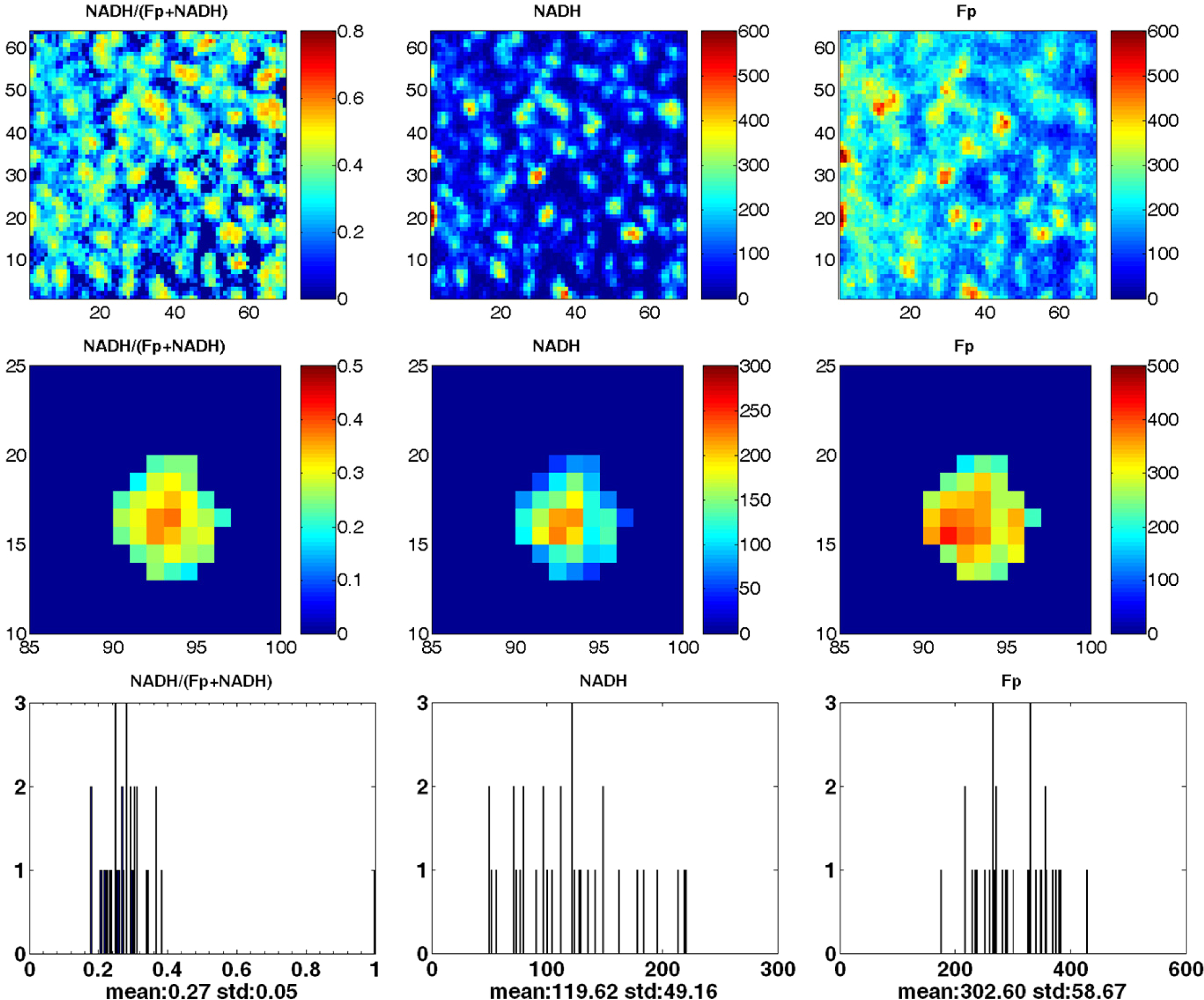
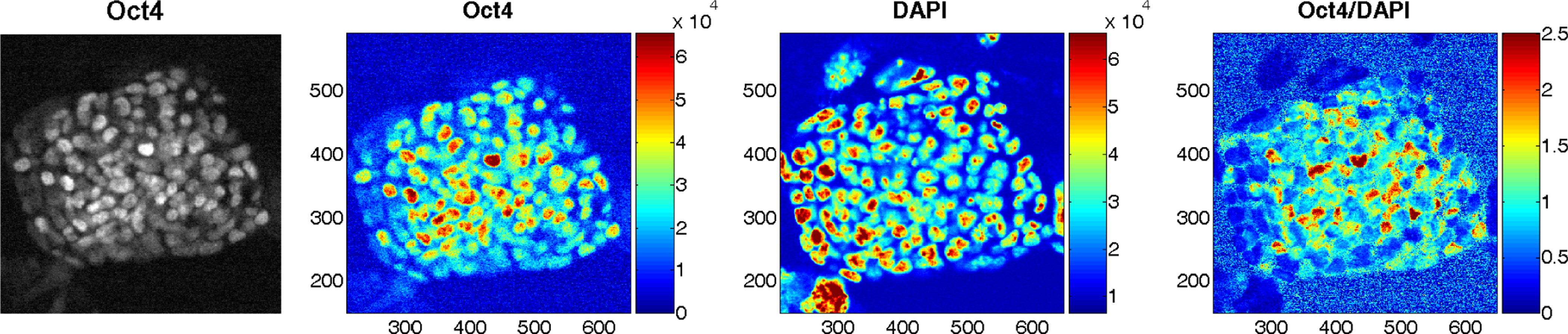
Redox state mediates embryonic stem cell (ESC) differentiation and thus offers an important complementary approach to understanding the pluripotency of stem cells. NADH redox ratio (NADH/(Fp + NADH)), where NADH is the reduced form of nicotinamide adenine dinucleotide and Fp is the oxidized flavoproteins, has been established as a sensitive indicator of mitochondrial redox state. In this paper, we report our redox imaging data on the mitochondrial redox state of mouse ESC (mESC) colonies and the implications thereof. The low-temperature NADH/Fp redox scanner was employed to image mESC colonies grown on a feeder layer of gamma-irradiated mouse embryonic fibroblasts (MEFs) on glass cover slips. The result showed significant heterogeneity in the mitochondrial redox state within individual mESC colonies (size: ∼200-440 μm), exhibiting a core with a more reduced state than the periphery. This more reduced state positively correlates with the expression pattern of Oct4, a well-established marker of pluripotency. Our observation is the first to show the heterogeneity in the mitochondrial redox state within a mESC colony, suggesting that mitochondrial redox state should be further investigated as a potential new biomarker for the stemness of embryonic stem cells.
Keywords: NADH; Redox imaging; flavoproteins; pluripotency; redox ratio.
Figures
References
-
- Evans MJ, Kaufman MH, “Establishment in culture of pluripotential cells from mouse embryos,” Nature 292, 154–156 (1981). - PubMed
-
- Thomson JA, Itskovitz-Eldor J, Shapiro SS, Waknitz MA, Swiergiel JJ, Marshall VS, Jones JM, “Embryonic stem cell lines derived from human blastocysts,” Science 282, 1145–1147 (1998). - PubMed
-
- Beddington RS, Robertson EJ, “An assessment of the developmental potential of embryonic stem cells in the midgestation mouse embryo,” Development 105, 733–737 (1989). - PubMed
-
- Rossant J, “Stem cells and lineage development in the mammalian blastocyst,” Reprod. Fertil. Dev 19, 111–118 (2007). - PubMed
Grants and funding
LinkOut - more resources
Full Text Sources