. 2021 Mar 30;8(10):2468-2485.
doi: 10.1039/d1qi00211b.

Development of a cobalt(iii)-based ponatinib prodrug system

Affiliations

Development of a cobalt(iii)-based ponatinib prodrug system

Marlene Mathuber et al. Inorg Chem Front. .

Abstract

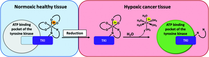
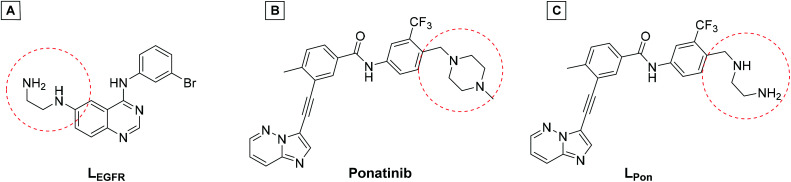
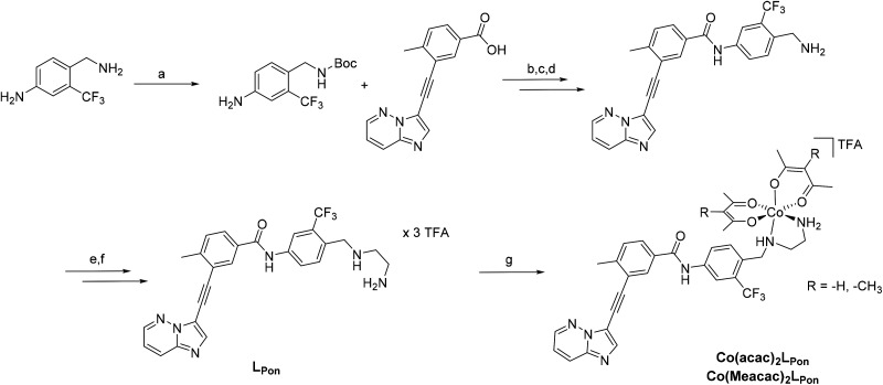
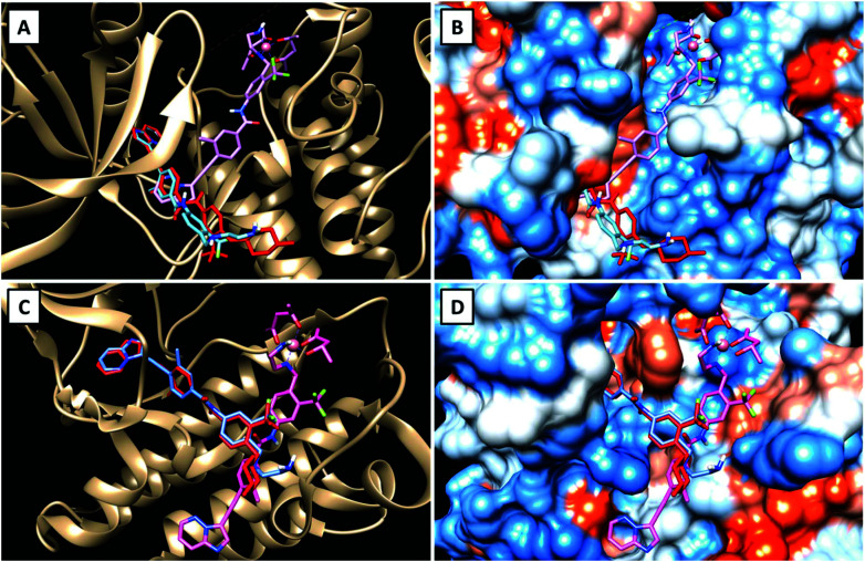
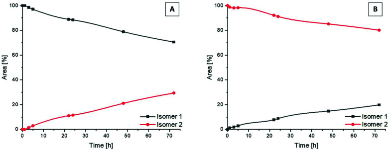
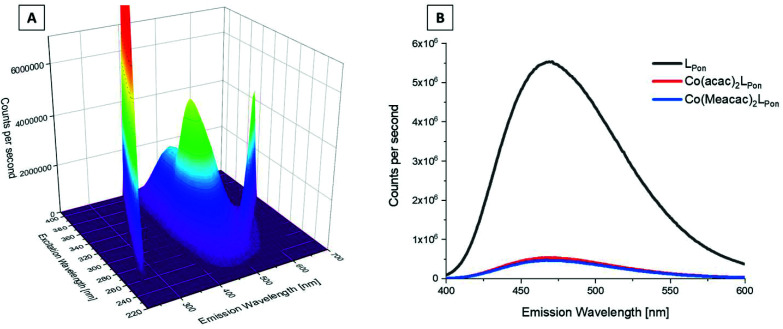
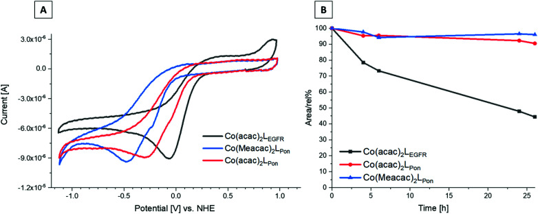
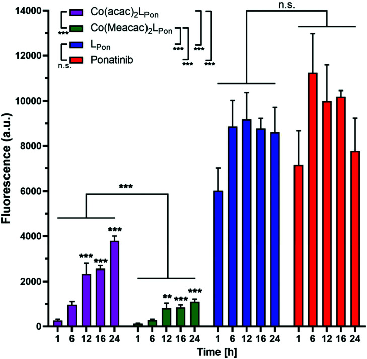
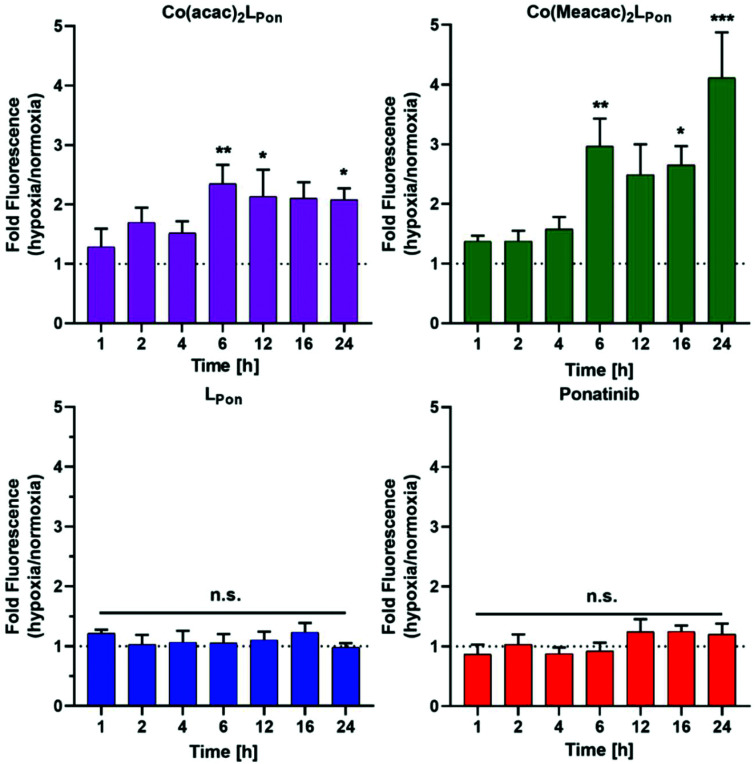
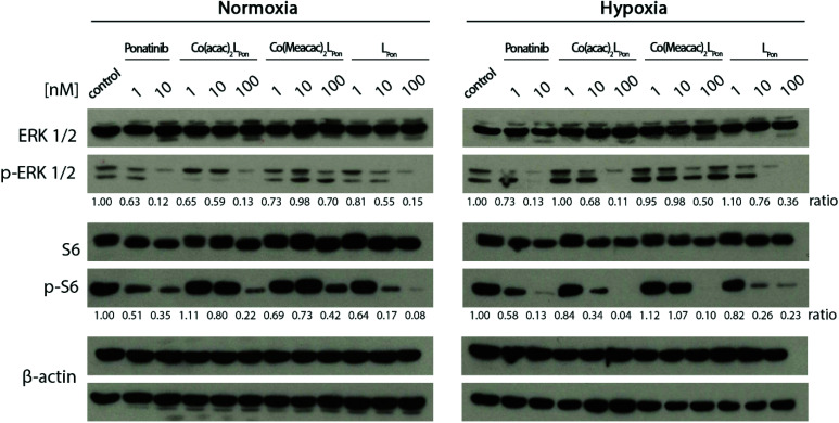
Receptor tyrosine kinase inhibitors have become a central part of modern targeted cancer therapy. However, their curative potential is distinctly limited by both rapid resistance development and severe adverse effects. Consequently, tumor-specific drug activation based on prodrug designs, exploiting tumor-specific properties such as hypoxic oxygen conditions, is a feasible strategy to widen the therapeutic window. After proof-of-principal molecular docking studies, we have synthesized two cobalt(iii) complexes using a derivative of the clinically approved Abelson (ABL) kinase and fibroblast growth factor receptor (FGFR) inhibitor ponatinib. Acetylacetone (acac) or methylacetylacetone (Meacac) have been used as ancillary ligands to modulate the reduction potential. The ponatinib derivative, characterized by an ethylenediamine moiety instead of the piperazine ring, exhibited comparable cell-free target kinase inhibition potency. Hypoxia-dependent release of the ligand from the cobalt(iii) complexes was proven by changed fluorescence properties, enhanced downstream signaling inhibition and increased in vitro anticancer activity in BCR-ABL- and FGFR-driven cancer models. Respective tumor-inhibiting in vivo effects in the BCR-ABL-driven K-562 leukemia model were restricted to the cobalt(iii) complex with the higher reduction potential and confirmed in a FGFR-driven urothelial carcinoma xenograft model. Summarizing, we here present for the first time hypoxia-activatable prodrugs of the clinically approved tyrosine kinase inhibitor ponatinib and a correlation of the in vivo activity with their reduction potential.


Conflict of interest statement

There are no conflicts to declare.

Figures

Fig. 1
Fig. 1. Proposed mode of action of hypoxia-activated cobalt(iii) prodrugs. Normoxic, healthy tissue (left side): The prodrug is inactive. Due to its bulkiness, the cobalt(iii) complex does not fit into the ATP-binding pocket of the respective tyrosine kinase. Hypoxic cancer tissue (right side): The drug is active. In the hypoxic tissue of the tumor, the cobalt(iii) complex is irreversibly reduced and the TKI ligand subsequently released able to inhibit the signaling processes of the target tyrosine kinases.
Fig. 2
Fig. 2. (A) Chemical structure of the previously synthesized EGFR inhibitor ligand (LEGFR) with a direct attachment of the ethylenediamine moiety to the aromatic ring system. (B) The clinically approved ponatinib (Iclusig®). (C) Newly developed ponatinib derivative (LPon), where the metal-chelating ethylenediamine moiety is separated from the aromatic ring system.
Scheme 1
Scheme 1. Synthesis of LPon and its cobalt(iii) complexes Co(acac)2LPon and Co(Meacac)2LPon. Reagents and conditions: (a) Di-tert-butyl dicarbonat, THF, 81%; (b) (COCl)2, abs. Toluol, NMM, DMAP, 68%; (c) 4 N HCl in dioxan/H2O, 95%; (d) NaHCO3 in EtOAc, 94%; (e) N-Boc-2-aminoacetaldehyde, sodium cyanoborhydride, abs. THF, molecular sieves (3–4 Å), 50%; (f) TFA in dichloromethane (ratio 1 : 1), 40%; (g) in situ deprotonation with NaOH, Na[Co(acac)2(NO2)2] or Na[Co(Meacac)2(NO2)2], activated charcoal in MeOH, 36% for Co(acac)2LPon and 31% for Co(Meacac)2LPon.
Fig. 3
Fig. 3. Visualizations of the best docking poses of LPon and Co(acac)2LPon in comparison to ponatinib with FGFR1 (A and B) (PDB ID: 4V04) as well as ABL1 (C and D) (PDB ID: 4WA9). Ponatinib is shown in red, LPon in blue and Co(acac)2LPon in pink.
Fig. 4
Fig. 4. Kinetic behavior of the isomers of Co(acac)2LPon incubated in PBS at 37 °C (pH 7.4, 10 mM) and monitored by HPLC. Depicted is the conversion from pure isomer 1 (A) and pure isomer 2 (B) into the respective mixtures over a period of 72 h.
Fig. 5
Fig. 5. Fluorescence properties of investigated ponatinib derivatives. (A) 3D full excitation–emission landscape of LPon (Rayleigh scattering of 1st and 2nd order appear as diagonal ridges). (B) Fluorescence emission spectra at λex = 320 nm of LPon, Co(acac)2LPon, and Co(Meacac)2LPon. Measurements were performed in PBS at pH = 7.40 [conc. ligand/complex = 15 μM; T = 25.0 °C].
Fig. 6
Fig. 6. (A) Cyclic voltammograms of Co(acac)2LPon, Co(Meacac)2LPon and Co(acac)2LEGFR in DMF (1.5 mM complex, I = 0.2 M [n-Bu4N][BF4], scan rate of 100 mV s−1, 25.0 °C). (B) Stability measurements of Co(acac)2LPon, Co(Meacac)2LPon and Co(acac)2LEGFR incubated in pure FCS at 37 °C (pH 7.4, 150 mM phosphate buffer) analyzed by HPLC-MS over a time period of 26 h. The y-axis shows the relative ratio of the integrated peak areas of the intact complex over time (in percent) compared to the area at the starting point (0 h).
Fig. 7
Fig. 7. Fluorescence-based evaluation of LPon release from Co(acac)2LPon and Co(Meacac)2LPon in K-562 cells under normoxic cell culture conditions by flow cytometry. Cells were incubated with 10 μM of the respective compounds for the indicated exposure times and mean fluorescence intensities were determined by flow cytometry (BD LSRFortessa X-20, pacific blue filter). Fluorescence intensities were normalized by subtracting the auto fluorescence of untreated cells. Data are given in arbitrary units (a.u.) as means ± SEM of five independent experiments. Statistical significance was calculated by two-way ANOVA with p < 0.05(*); <0.01 (**); <0.001 (***).
Fig. 8
Fig. 8. Impact of hypoxia on cell-associated LPon-fluorescence from Co(acac)2LPon and Co(Meacac)2LPon. K-562 cells were incubated with 10 μM of compounds for the indicated time points under hypoxic and normoxic conditions and fluorescence intensities were determined by flow cytometry (BD LSRFortessa X-20, pacific blue filter). Fluorescence intensities were determined as described under Fig. 7 and mean fluorescence intensities under hypoxic cell culture conditions were normalized to the respective normoxic conditions. Data are given as means ± SEM of five independent experiments. Statistical significance was calculated by one-way ANOVA with Dunnett multiple comparison test with p < 0.05(*); <0.01 (**); <0.001 (***).
Fig. 9
Fig. 9. Anticancer activity of Co(acac)2LPon, Co(Meacac)2LPon, LPon and ponatinib against human cancer cell models under normoxic (N) and hypoxic (H) conditions. (A) BCR-ABL-positive K-562 leukemic cell viability was measured by MTT vitality assay and (B) by luminescence assay based on ATP quantification (CellTiter-Glo) after 72 h. (C) Clonogenic cell growth of the FGFR3-driven UM-UC-14 urothelial cancer cell model determined by colony formation assay after 5 d of incubation. Data are given as means ± SD of one representative experiment performed in triplicates. Statistical significance was calculated by two-way ANOVA with Sidak multiple comparison test with p < 0.05(*); <0.01 (**); <0.001 (***).
Fig. 10
Fig. 10. Impact of the cobalt(iii) complexes on the ERK1/2 and S6 under normoxic and hypoxic conditions. K-562 cells were treated with the compounds, after 12 h cell lysates were prepared and protein expression as well as phosphorylation levels of downstream pathways (p-ERK1/2 and p-S6) analyzed by western blotting. One representative experiment out of three is shown. The ratio of phosphorylated to total protein is given between the respective lanes.
Fig. 11
Fig. 11. In vivo anticancer activity of the investigated cobalt(iii) complexes. (A) BCR-ABL-driven leukemic K-562 cells or (B) FGFR3-driven urothelial UM-UC-14 cells were injected s.c. into the right flank of male CB17/SCID mice (n = 4 animals per experimental group). When tumors were measurable (day 5 and day 7, respectively) compounds (10 mg kg−1 i.p.) were applied as indicated (black arrows). Tumor sizes were evaluated by caliper measurement. Data are given as means ± SEM. Statistical significance was calculated by two-way ANOVA with Sidak multiple comparison test with p < 0.05(*); <0.01 (**).

Similar articles

Cited by

References

    1. Lemmon M. A. Schlessinger J. Cell signaling by receptor tyrosine kinases. Cell. 2010;141:1117–1134. - PMC - PubMed
    1. Du Z. Lovly C. M. Mechanisms of receptor tyrosine kinase activation in cancer. Mol. Cancer. 2018;17:58. - PMC - PubMed
    1. Wu P. Nielsen T. E. Clausen M. H. FDA-approved small-molecule kinase inhibitors. Trends Pharmacol. Sci. 2015;36:422–439. - PubMed
    1. Chiavenna S. M. Jaworski J. P. Vendrell A. State of the art in anti-cancer mAbs. J. Biomed. Sci. 2017;24:1–12. - PMC - PubMed
    1. Huang W.-S. Metcalf C. A. Sundaramoorthi R. Wang Y. Zou D. Thomas R. M. Zhu X. Cai L. Wen D. Liu S. Discovery of 3-[2-(imidazo [1, 2-b] pyridazin-3-yl) ethynyl]-4-methyl-N-{4-[(4-methylpiperazin-1-yl) methyl]-3-(trifluoromethyl) phenyl} benzamide (AP24534), a potent, orally active pan-inhibitor of breakpoint cluster region-abelson (BCR-ABL) kinase including the T315I gatekeeper mutant. J. Med. Chem. 2010;53:4701–4719. - PubMed

LinkOut - more resources