. 2021 May 20:8:451-465.
doi: 10.2147/JHC.S303588. eCollection 2021.

The Crosstalk Between Cancer Cells and Neutrophils Enhances Hepatocellular Carcinoma Metastasis via Neutrophil Extracellular Traps-Associated Cathepsin G Component: A Potential Therapeutic Target

Affiliations

The Crosstalk Between Cancer Cells and Neutrophils Enhances Hepatocellular Carcinoma Metastasis via Neutrophil Extracellular Traps-Associated Cathepsin G Component: A Potential Therapeutic Target

Xiangqian Guan et al. J Hepatocell Carcinoma. .

Abstract

Background: Emerging evidences have highlighted the roles of neutrophils, as the major host microenvironment component, in the development of hepatocellular carcinoma (HCC). Neutrophils extracellular traps (NETs) produced in the infection can strengthen the behavior of cancer metastasis. Here, we investigated the roles of NETs in HCC metastasis and further explore the underlying mechanism of how NETs interact with cancer.

Methods: The neutrophils were isolated from whole blood of HCC patients and used to evaluate the formation of NETs. NET markers were detected in tissue samples, plasma and cell climbing slice. Mouse models were used to evaluate the roles of NETs in HCC metastasis in vivo, and the corresponding mechanisms were explored using in vivo and in vitro assays.

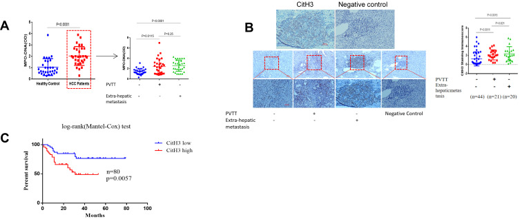
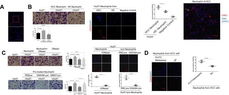
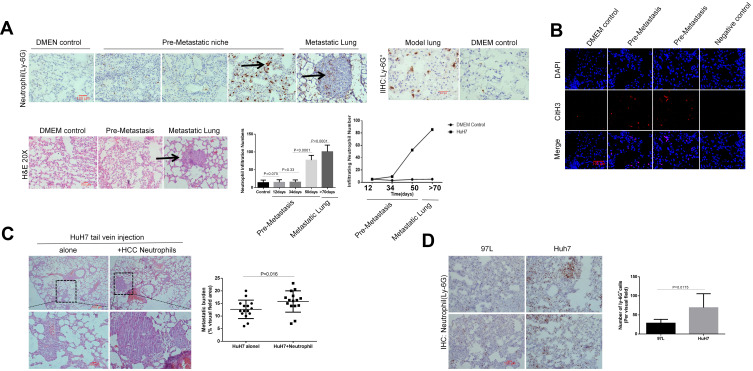
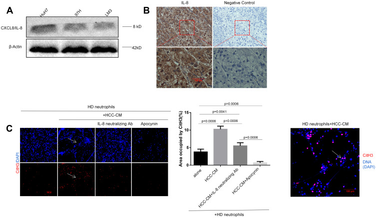
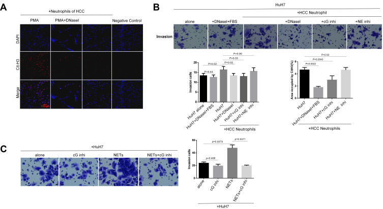
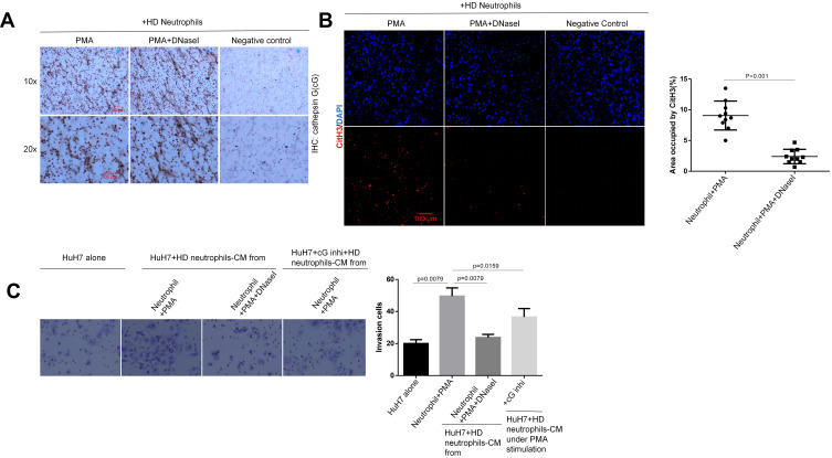
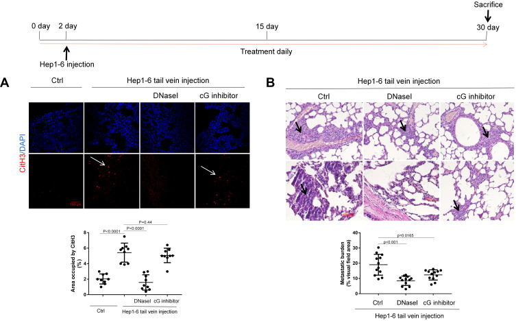
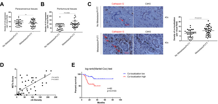
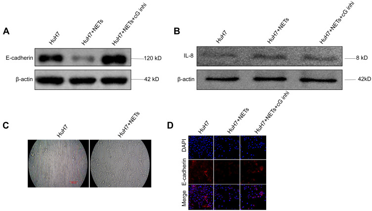
Results: An increase in the release of NETs in patients with HCC, particularly those with portal vein tumor thrombosis (PVTT). The presence of NETs in HCC tumor tissues closely correlated with a poor prognosis. Functionally, the invasion ability of HCC cells was enhanced by co-culture with HCC neutrophils, through NETs formation, while the neutrophils from a healthy donor (HD) exhibited the inhibition of the invasion ability. Furthermore, we observed an enhanced ability of forming NETs in neutrophils from HCC patients in vitro, especially patients with PVTT or extra-hepatic metastasis. An in-vivo animal study demonstrated that neutrophils of HCC facilitated the metastatic behavior towards the lung. The further mechanistic investigation unveiled that HCC cells-derived cytokine IL-8 triggered NETs formation in an NADPH oxidase-dependent manner, and NETs-associated cathepsin G (cG) promoted HCC metastasis in vitro as well as vivo. Clinically, the expression of the cG protein in tumor tissues displayed a close correlation with the disease prognosis of HCC patients.

Conclusion: Our findings implicated that the induction of NETs by HCC cells is a critical metastasis-supporting cancer-host interaction and that NETs may serve as an immune-based potential therapeutic target against HCC progression.

Keywords: E-cadherin; IL-8; NADPH; cathepsin G; hepatocellular carcinoma; metastasis; neutrophils extracellular traps.


Conflict of interest statement

The authors reported no conflicts of interest for this work.

Figures

Figure 1
Figure 1
Increased NETs marker in HCC patients. (A) The Levels of MPO-DNA in HCC or healthy control serum samples were examined by the capture ELISA assay (n=90). The red dotted box points to HCC patients, indicating that MPO-DNA levels were particularly elevated in either PVTT or extra-hepatic metastasis among HCC patients. (B) The deposition of NETs-CitH3 in 85 HCC tissue samples were examined by the immunohistochemistry assay. NETs-CitH3 level elevated in HCC patients tissue with portal vein tumor thrombosis/extra-hepatic metastasis(n=41), compared to those without metastasis(n=44). Representative images of CitH3 in human tissues were shown. Scare bar: 100um. (C) Presence of NETs-CitH3 in HCC tissues correlated with disease prognosis. Log-rank (Mantel-Cox) disease-free survival curves of HCC patients was used basing on tissue CitH3 immunoscore.
Figure 2
Figure 2
Neutrophil of HCC facilitates HCC cells invasion through NETs formation in vitro. (A) DAPI staining showed the purity of the neutrophils isolated from human peripheral whole blood. Scare bar: 100um. (B) The representative images of transwell assay for the effects of neutrophils on HCC cells invasion were shown. The cells were counted under a microscope in twenty randomly selected fields. NETs were identified by CitH3 component. The expression of NETs-CitH3 was detected by immunofluorescence staining on coverslip. The white arrow points to the expression and presence of NETs-CitH3. Scare bar: 100um. (C) The representative images of transwell assay for the effects of DNaseI or PAD4 inhibitor GSK484 on neutrophils-induced HCC cells invasion were shown. The expression of NETs-CitH3 was detected by immunofluorescence staining on coverslip. Neutrophils of HCC was pre-treated with GSK484 at 10uM for 4h before co-culture. 15µL DNaseI was added into per 500µL culture medium in lower chamber. Scare bar: 100um. (D) The expression of NETs-CitH3 was detected by immunofluorescence staining for neutrophils from HCC patients with metastasis or without.Scare bar: 100µm.
Figure 3
Figure 3
Neutrophils of HCC enhances metastasis burden in vivo. (A) HuH7 cells were injected into nude mice via tail vein. At various time points (12, 34, 50days) after HCC cells injection, the mice was killed and examined the density of ly-6G+cells infiltration. Ly-6G+cells in nude mice lung were shown by immunohistochemistry staining with lung frozen section (n=7 each). Representative images of ly-6G+ neutrophils in the lung were shown. The black arrows point to Ly-6G+ neutrophils (IHC staining) and metastasis (H&E staining). Scare bar: 100µm. (B) NETs-CitH3 were detected by immunofluorescence staining with lung frozen section. Representative images of CitH3 in the lung were shown (n=7 each). Scare bar: 100µm. (C) HuH7 cells were co-injected with HCC neutrophils (10:1) into nude mice (n=7 each). The metastatic burden in each group were calculated as the percentage of area occupied by metastatic tumor on each lung section, determined using Image J software. Representative images of metastatic foci in lung were shown. Scare bar: 100µm. (D) Ly-6G+ cells were detected by immunohistochemistry with frozen lung section after HuH7 or 97L injection (n=5each). Representative images were shown. Scare bar: 100µm.
Figure 4
Figure 4
HCC cells-derived CXCL8/IL-8 triggers TAN towards NETs formation through NADPH pathway. (A) The protein expression of IL-8 was detected by Western blot assay in three HCC cell lines. Experiment was repeated three times. (B) The expression of IL-8 in HCC tissues was examined by the immunohistochemistry (n=30). A representative image was shown. Scare bar: 100um. (C) Co-incubation of HD neutrophils (2.5x10⁶/mL) with the conditioned medium of HCC cells (HCC-CM) (1:2) was performed to explore the effect of IL-8 on HCC-CM-induced NETs formation. The expression of CitH3 was detected by immunofluorescence staining on coverslip for the identification of NETs formation. Representative images were shown. IL-8 neutralizing antibody was added into the culture medium at the concentration of 5µg/mL. NOX inhibitor apocynin was added into medium at the final concentration of 10uM. Experiment was repeated three times. Representative images were shown. The white arrows point to the presence of NETs-CitH3. Scare bar: 100µm.
Figure 5
Figure 5
NETs-associated cathepsin G promotes HCC cells invasion. (A) Neutrophils generated NETs in response to PMA. PMA was co-incubated with neutrophils at a final concentration of 500nM for the preparation of NETs. DNaseI abolished the process of PMA-induced NETs formation. The expression of CitH3 was detected by immunofluorescence staining on coverslip for the identification of NETs formation. Representative images were shown. Scare bar: 100µm. (B) cG inhibitor reduced NETs extension and neutrophils-induced HCC cells invasion. NE inhibitor had no significant effect on NETs formation and neutrophils-induced invasion. Representative images were shown. cG inhibitor was added into culture medium at a final concentration of 2µM, and NE inhibitor was added into culture medium at a final concentration of 10uM. Experiment was repeated three times. (C) cG inhibitor abolished NETs-stimulated HCC cells invasion. cG inhibitor was added into culture medium at the final concentration of 2µM. Control group was set with DMEM culture medium. Representative images were shown. Experiment was repeated three times.
Figure 6
Figure 6
Co-localization of cG and CitH3 is necessary to promote invasion in vitro. (A) The cathepsin G expression of HD neutrophils on coverslip were shown by immunohistochemistry staining. Representative images were shown. Scare bar: 100µm. (B) The expression of CitH3 was detected by immunofluorescence staining on coverslip for the identification of NETs formation. Representative images were shown. Scare bar: 100µm. (C) Transwell assay was performed to detect the invasion-inducing capacity of HD neutrophils-CM under different treatment. cG inhibitor was added into culture medium at the final concentration of 2µM. Control group was set with DMEM culture medium. Representative images were shown. Experiment was repeated three times.
Figure 7
Figure 7
Inhibition of cG component alleviates lung metastasis in vivo. (A) The expression of NETs-CitH3 protein in model lung were shown by immunofluorescence (n=5 each). Representative images were shown. The white arrows point to the expression and presence of NETs-CitH3 in lung. Scare bar: 100µm. (B) Hep1-6 cells were injected into male C57BL/6J mice via tail vein (n=5 each), and the metastatic burden in each group were calculated as the percentage of area occupied by metastatic tumor on each lung tissue section, determined using Image J software. Representative images of metastatic foci were shown. The black arrows point to metastasis foci. Scare bar: 100µm.
Figure 8
Figure 8
Co-localization between cG and NETs-CitH3 in HCC tissues indicates disease prognosis. (A–C) The expression of cathepsin G and CitH3 protein in patients tissues were shown by immunohistochemistry staining with serial sections (n=60). Representative images were shown. The red arrows point to protein cathepsin G and black arrows point to protein CitH3 in serial section, indicating the co-localization of protein cathepsin G and CitH3 in tissue. Scare bar: 100µm. (D) Pearson correlation analysis of NETs-CitH3 and cG expression in HCC samples (n=60). (E) 60 non-metastasis/PVTT HCC patients with different level of cG and NETs for metastasis/PVTT cumulative incidence analysis, and Log rank test was used.
Figure 9
Figure 9
NETs-associated cathepsin G regulates the expression of E-cadherin in HCC cells in vitro. (A, B) Western blot analysis of E-cadherin and CXCL8/IL-8 level in HuH7 cells treated with NETs-CM. Cells were stimulated with NETs-CM for 20 hours. Experiment was repeated three times. (C) HuH7 (5x10⁵ cells) were co-cultivated with NETs-CM or cultivated alone to confluence for 20 hours, and the area depleted of tumor cells was quantified as percentage of the total area. Representative images were shown. Scare bar: 100µm. (D) E-cadherin expression of HuH7 cells layer was shown by immunofluorescence staining. Representative images were shown. Scare bar: 100um. Experiment was repeated three times.

Similar articles

Cited by

References

    1. Guo L, Chen G, Zhang W, et al. A high-risk luminal A dominant breast cancer subtype with increased mobility. Breast Cancer Res Treat. 2019;175(2):459–472. - PMC - PubMed
    1. Berger-Achituv S, Brinkmann V, Abed UA, et al. A proposed role for neutrophil extracellular traps in cancer immunoediting. Front Immunol. 2013;4:48. - PMC - PubMed
    1. Papayannopoulos V. Neutrophil extracellular traps in immunity and disease. Nat Rev Immunol. 2018;18(2):134–147. - PubMed
    1. Quail DF, Joyce JA. Microenvironmental regulation of tumor progression and metastasis. Nat Med. 2013;19(11):1423–1437. - PMC - PubMed
    1. Albrengues J, Shields MA, Ng D, et al. Neutrophil extracellular traps produced during inflammation awaken dormant cancer cells in mice. Science. 2018;361:6409. - PMC - PubMed