Maisonneuve injury with no fibula fracture: A case report
- PMID: 34046477
- PMCID: PMC8130071
- DOI: 10.12998/wjcc.v9.i15.3733
Maisonneuve injury with no fibula fracture: A case report
Abstract
Background: Ankle syndesmosis injury is difficult to diagnose accurately at the initial visit. Missed diagnosis or improper treatment can lead to chronic complications. Complete syndesmosis injury with a concomitant rupture of the interosseous membrane (IOM) is more unstable and severe. The relationship between this type of injury and Maisonneuve injury, in which the syndesmosis is also injured, has not been discussed in the literature previously.
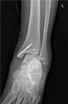
Case summary: A 16-year-old patient sustained left medial malleolar fracture, and the associated inferior tibiofibular syndesmotic instability was overlooked. After open reduction and internal fixation of the medial malleolar fracture, inferior tibiofibular syndesmosis diastasis with IOM rupture was detected by auxiliary imaging. Secondary surgical intervention was performed to reduce anatomically and fix with two trans-syndesmosis screws. Twelve weeks later, the screws were removed. At the 6-mo follow-up, the patient gained full range of motion of the ankle.
Conclusion: Complete syndesmosis injury with IOM rupture should be considered Maisonneuve-type injury. Open reduction and internal fixation could obtain good outcomes.
Keywords: Case report; Classification; Interosseous membrane; Maisonneuve; Stress test; Syndesmosis injury.
©The Author(s) 2021. Published by Baishideng Publishing Group Inc. All rights reserved.
Conflict of interest statement
Conflict-of-interest statement: The authors declare that they have no conflict of interest to report.
Figures


References
-
- Maisonneuve MJG. Recherches sur la fracture du péroné. Arch Gen Med. 1840;7:165–187.
-
- Stufkens SA, van den Bekerom MP, Doornberg JN, van Dijk CN, Kloen P. Evidence-based treatment of maisonneuve fractures. J Foot Ankle Surg. 2011;50:62–67. - PubMed
-
- Lock TR, Schaffer JJ, Manoli A 2nd. Maisonneuve fracture: case report of a missed diagnosis. Ann Emerg Med. 1987;16:805–807. - PubMed
-
- McCollum GA, van den Bekerom MP, Kerkhoffs GM, Calder JD, van Dijk CN. Syndesmosis and deltoid ligament injuries in the athlete. Knee Surg Sports Traumatol Arthrosc. 2013;21:1328–1337. - PubMed
-
- Tourné Y, Molinier F, Andrieu M, Porta J, Barbier G. Diagnosis and treatment of tibiofibular syndesmosis lesions. Orthop Traumatol Surg Res. 2019;105:S275–S286. - PubMed
Publication types
LinkOut - more resources
Full Text Sources
Other Literature Sources

