

Lymphangiomatosis associated with protein losing enteropathy: A case report
- PMID: 34046480
- PMCID: PMC8130063
- DOI: 10.12998/wjcc.v9.i15.3758
Lymphangiomatosis associated with protein losing enteropathy: A case report
Abstract
Background: Lymphangiomatosis is a multisystem disorder that is rarely localized to the gastrointestinal tract. Lymphangiomatosis usually has no specific clinical presentation and is easily misdiagnosed. A case report and review of the literature on lymphangiomatosis associated with protein-losing enteropathy will help to improve the overall understanding of this disease.
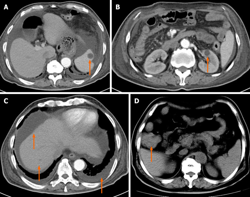
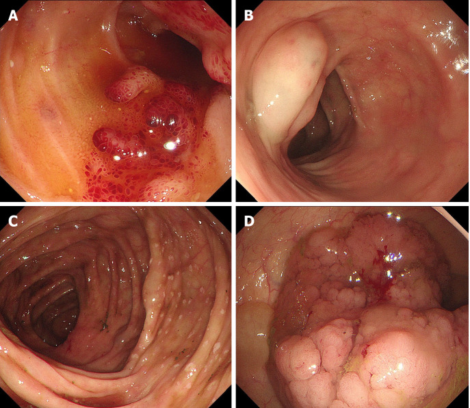
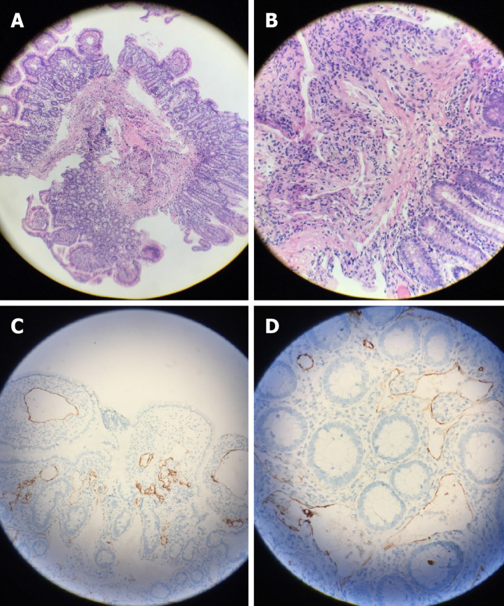
Case summary: We report a case of lymphangiomatosis of the bowel and other solid organs. A 78-year-old man presented with recurrent bowel bleeding and protein-losing enteropathy, as well as cystic lesions in the spleen, liver, and kidney. Imaging examinations revealed many cystic lesions on the spleen, liver, kidney, and thickened wall of the ascending colon, as well as pleural effusion and ascites. Colonoscopy revealed a strawberry mucosa, variable spontaneous bleeding, and surface erosion located in the terminal ileum. Several cystic masses with a translucent and smooth surface as well as diffuse white spots were located in the colon. A laterally spreading tumor (LST) was located in the ascending colon. Pathology indicated highly differentiated adenocarcinoma (LST) and lymphangiomatoid dilation, and D2-40 was positive. The final diagnosis was lymphangiomatosis. The patient underwent surgery for LST and then was administered thalidomide 75-150 mg/d. His condition, however, did not improve. He eventually died 6 mo after the initial diagnosis.
Conclusion: Lymphangiomatosis usually occurs diffusely and can involve many organs, such as the spleen, kidney, liver, lung, mesentery, and bowel. Recurrent bowel bleeding or protein-losing enteropathy is an important indicator that should alert clinicians about the possibility of this disease when it afflicts the bowel. Doctors should improve the medical understanding of lymphangiomatosis.
Keywords: Case report; Colonic neoplasms; Gastrointestinal hemorrhage; Lymphangioma; Protein-losing enteropathies; Small intestine.
©The Author(s) 2021. Published by Baishideng Publishing Group Inc. All rights reserved.
Conflict of interest statement
Conflict-of-interest statement: The authors declare that they have no conflicts of interest to report.
Figures
References
-
- Valakada J, Madhusudhan KS, Ranjan G, Garg PK, Sharma R, Gupta AK. Abdominal Lymphangiomatosis With Intestinal Lymphangiectasia Diagnosed by Magnetic Resonance Lymphangiography: A Case Report. Curr Probl Diagn Radiol. 2018;47:200–202. - PubMed
-
- Glöckler M, Severin T, Arnold R, Greiner P, Schwab KO, Uhl M, Schlensak C, Rössler J, Dittrich S. First description of three patients with multifocal lymphangiomatosis and protein-losing enteropathy following palliation of complex congenital heart disease with total cavo-pulmonary connection. Pediatr Cardiol. 2008;29:771–774. - PubMed
Publication types
LinkOut - more resources
Full Text Sources
Other Literature Sources
