. 2021 May 8:2021:9966344.
doi: 10.1155/2021/9966344. eCollection 2021.

Effects and Anti-rotation Stabilization of the Non-bridging External Fixation for Pronation-Abduction Stage III Ankle Fracture: A Cadaveric Study

Affiliations

Effects and Anti-rotation Stabilization of the Non-bridging External Fixation for Pronation-Abduction Stage III Ankle Fracture: A Cadaveric Study

Yili Chen et al. Biomed Res Int. .

Abstract

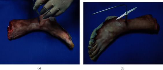
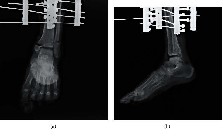
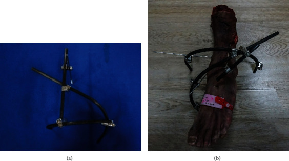
Objective: This study is aimed at providing a nonbridging external fixation technique with pinning fixation for the pronation-abduction stage III ankle fracture. The secondary purpose was to evaluate its effect on anatomic reduction and fracture fragment stability against cadaveric models' rotation.

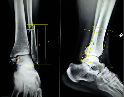
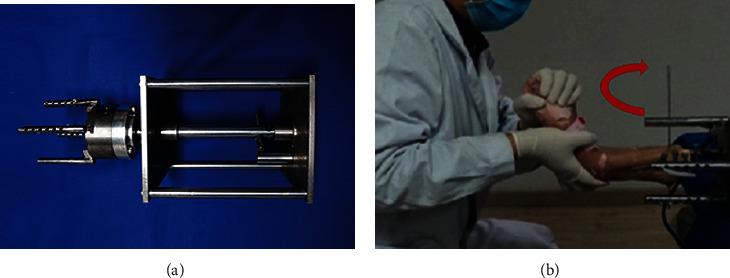
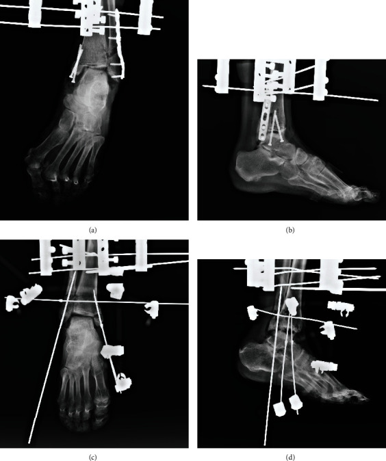
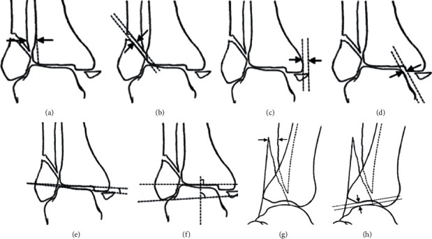
Method: A paired design study was conducted using 14 pairs of the cadaveric model which had been modeled for pronation-abduction stage III ankle fracture. One fracture model from each pair was randomly allocated to receive an open reduction and internal fixation, while the other was reduced and stabilized with the external fixation technique. After the surgery, the antirotational stability tests were performed with external rotation torques of 10 nm, 15 nm, and 20 nm applied, respectively. The postoperation reduction rate and ankle parameters were recorded in anteroposterior and lateral radiographs before and after the antirotational stability experiment.

Result: The outcomes were assessed according to Burwell-Charnley's radiographic criteria of reduction. It showed no statistically significant differences in reduction between the two groups (P < 0.05). The displacement of lateral fragment following a reduction in the external fixation group was significantly larger than that of the internal fixation group (3.14 ± 0.56 vs. 1.49 ± 0.39, P < 0.05). After applying rotational torques of 10 nm, 15 nm, and 20 nm, the results of other parameters showed no significant differences between the two groups.

Conclusion: This nonbridging external fixation method with pin fixation of fracture fragments might have the same effect as that of internal fixation on the reduction rate of pronation-abduction stage III ankle fracture.


Conflict of interest statement

It is to claim that there is no conflict of interest with any other individuals or government in the manuscript.

Figures

Figure 1
Figure 1
The osteotomy was to draw fracture lines on bone by using the drill bit guider. (a) The medial side. (b) The lateral side.
Figure 2
Figure 2
The fracture line of the medial and lateral malleoli obtained from the typical PAB stage III ankle plain radiographs using Digitizer 4.2.6.0. (i) Anteroposterior ankle view: (a) the lateral height of the lateral malleolus; (b) the medial height of the lateral malleolus; (c) the medial height of the medial malleolus; (d) the lateral height of the medial malleolus. (ii) Lateral ankle view: (e, f) the measurement of the lateral malleoli, which could indicate the fracture line trend.
Figure 3
Figure 3
The specimens were rigidly fixed to a mechanical measuring and specimen fixating machine, which was equipped with a force transducer. (a) The mechanical measuring and specimen fixating device. (b) The researcher held the heel with the right hand and held the metatarsophalangeal with the left hand, disposing the ankle in a pronation position and applying abduction force on the talus.
Figure 4
Figure 4
Radiographs were taken after modeling for the verification. A typical model's radiograph. (a) The anteroposterior view. (b) The lateral view.
Figure 5
Figure 5
(a) The nonbridging external fixator was constructed by a semicircle rod, an arc-shaped one, and a straight one. (b) The external fixator was installed on a PA stage III ankle fracture.
Figure 6
Figure 6
The anteroposterior and lateral radiographs were taken after fixation. (a, b) The internal fixation. (c, d) The nonbridging external fixation.
Figure 7
Figure 7
Illustrations of reduction evaluation parameters. AP radiograph: (a) the width of syndesmosis; (b) the lateral displacements (LMD); (c) the displacement of the medial malleolar fragment (MMD); (d) the distance of medial clear space (MCS); (e) talar tilt angle (TTA); (f) talocrural angle (TCA). LAT radiograph: (g) the lateral displacements (LMD); (h) the anterior talar translation (ATT). This picture is partially referenced by the citations [31, 32].

References

    1. Elsoe R., Ostgaard S. E., Larsen P. Population-based epidemiology of 9767 ankle fractures. Foot and Ankle Surgery. 2018;24(1):34–39. doi: 10.1016/j.fas.2016.11.002. - DOI - PubMed
    1. Thur C. K., Edgren G., Jansson K.-Å., Wretenberg P. Epidemiology of adult ankle fractures in Sweden between 1987 and 2004. Acta Orthopaedica. 2012;83(3):276–281. doi: 10.3109/17453674.2012.672091. - DOI - PMC - PubMed
    1. Juto H., Nilsson H., Morberg P. Epidemiology of adult ankle fractures: 1756 cases identified in Norrbotten County during 2009-2013 and classified according to AO/OTA. BMC Musculoskeletal Disorders. 2018;19(1) doi: 10.1186/s12891-018-2326-x. - DOI - PMC - PubMed
    1. Tartaglione J. P., Rosenbaum A. J., Abousayed M., DiPreta J. A. Classifications in brief: Lauge-Hansen classification of ankle fractures. Clinical Orthopaedics and Related Research®. 2015;473(10):3323–3328. doi: 10.1007/s11999-015-4306-x. - DOI - PMC - PubMed
    1. Siegel J., Tornetta P. Extraperiosteal plating of pronation-abduction ankle fractures. Journal of Bone & Joint Surgery. 2007;89(2) doi: 10.2106/jbjs.e.00987. - DOI - PubMed

LinkOut - more resources