Review
. 2021 Apr 14:10:38.
doi: 10.12703/r/10-38. eCollection 2021.

Neural crest multipotency and specification: power and limits of single cell transcriptomic approaches

Affiliations
Review

Neural crest multipotency and specification: power and limits of single cell transcriptomic approaches

Kristin B Artinger et al. Fac Rev. .

Abstract

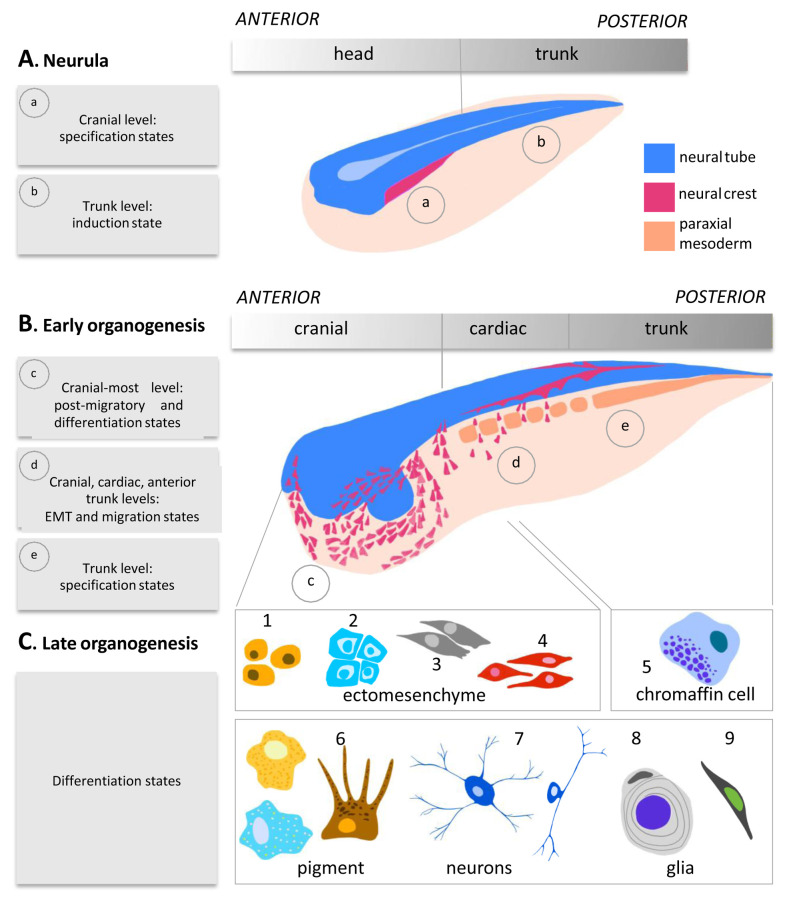
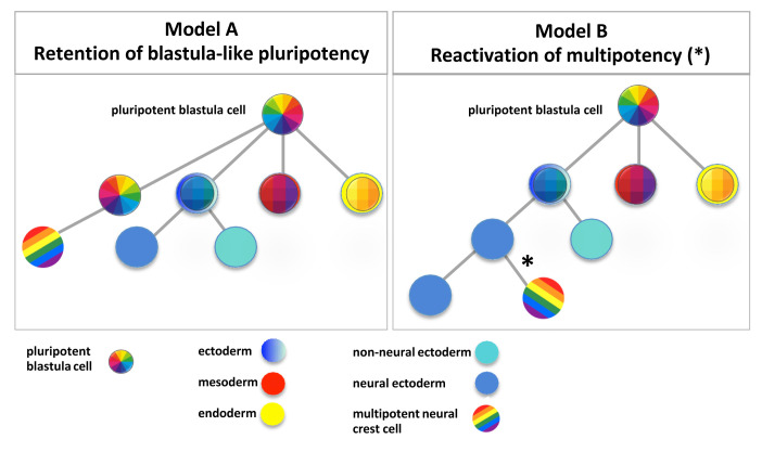
The neural crest is a unique population of multipotent cells forming in vertebrate embryos. Their vast cell fate potential enables the generation of a diverse array of differentiated cell types in vivo. These include, among others, connective tissue, cartilage and bone of the face and skull, neurons and glia of the peripheral nervous system (including enteric nervous system), and melanocytes. Following migration, these derivatives extensively populate multiple germ layers. Within the competent neural border ectoderm, an area located at the junction between the neural and non-neural ectoderm during embryonic development, neural crest cells form in response to a series of inductive secreted cues including BMP, Wnt, and FGF signals. As cells become progressively specified, they express transcriptional modules conducive with their stage of fate determination or cell state. Those sequential states include the neural border state, the premigratory neural crest state, the epithelium-to-mesenchyme transitional state, and the migratory state to end with post-migratory and differentiation states. However, despite the extensive knowledge accumulated over 150 years of neural crest biology, many key questions remain open, in particular the timing of neural crest lineage determination, the control of potency during early developmental stages, and the lineage relationships between different subpopulations of neural crest cells. In this review, we discuss the recent advances in understanding early neural crest formation using cutting-edge high-throughput single cell sequencing approaches. We will discuss how this new transcriptomic data, from 2017 to 2021, has advanced our knowledge of the steps in neural crest cell lineage commitment and specification, the mechanisms driving multipotency, and diversification. We will then discuss the questions that remain to be resolved and how these approaches may continue to unveil the biology of these fascinating cells.

Keywords: fate specification; lineage specification; multipotency; neural crest; pluripotency; single cell transcriptomics.


Conflict of interest statement

The authors declare that they have no competing interests.No competing interests were disclosed.No competing interests were disclosed.No competing interests were disclosed.

Figures

Figure 1.
Figure 1.. Neural crest states over developmental time.
The steps of neural crest development during neurulation and organogenesis are schematised. A. During late gastrulation stage (not shown) and neurulation stage, neural crest cells are specified in an anterior-to-posterior wave, with cranial neural crest cells being more mature, e.g. being at specification state (a), while in the trunk, neural crest early induction steps are ongoing (b). B. From the end of neurulation to late organogenesis stages, neural crest cells undergo epithelial-to-mesenchyme transition (EMT), migrate, and differentiate. As earlier on, in a given embryo, many neural crest states co-exist. For example, in an early tadpole, anteriorly, cranial neural crest cells finish migration and initiate differentiation (c) while vagal neural crest cells (which include cardiac neural crest cells, at the levels of somite 1 to somite 3–4 in the chick embryo and prospective enteric neural crest cells at the levels of somite 3–8 in chick embryos) undergo EMT and early migration steps (d). In the same embryo, posteriorly, trunk neural crest is being specified dorsal to the neural tube (e). C. Last, cranial, cardiac, posterior vagal, and trunk neural crest cells differentiate into specific and common derivatives. Cranial and cardiac neural crest form ectomesenchyme: cranial cells form bone (1), cartilage (2), smooth muscle, and mesenchyme (3), and cardiac neural crest cells contribute to the outflow tract of the heart (4). Trunk-specific fates consist mainly of chromaffin cells in the adrenal medulla (5). At all levels of the anterior–posterior axis, neural crest cells generate pigment cells including melanocytes, xantophores, and iridophores (6), various types of neurons of the sensory, autonomous, and enteric nervous systems (7), Schwann cells and enteric glia (8), and Schwann cell progenitors (9). Neural tissue is depicted in blue, neural crest cells in magenta, and paraxial mesoderm in orange.
Figure 2.
Figure 2.. Proposed models of neural crest pluri/multipotency.
Two models are proposed to explain neural crest multipotency. A In this model, the neural crest lineage shares and retains a specific blastula-type gene signature of pluripotency (rainbow quadrants) while the three other germ layers are segregated into ectoderm (blue), mesoderm (red), or endoderm (yellow). Further subdivision of the ectoderm into neuroectoderm (sky blue) and non-neural ectoderm (turquoise) is indicated. B. In this model, the neural crest lineage re-activates (*) pluri/multipotency genes at the neural border (rainbow stripes) after neural lineage segregation (blue, red, and yellow as above).

Similar articles

Cited by

References

    1. Kumar P, Tan Y, Cahan P: Understanding development and stem cells using single cell-based analyses of gene expression. Development. 2017; 144(1): 17–32. 10.1242/dev.133058 - DOI - PMC - PubMed
    2. Faculty Opinions Recommendation

    1. Mitiku N, Baker JC: Genomic analysis of gastrulation and organogenesis in the mouse. Dev Cell. 2007; 13(6): 897–907. 10.1016/j.devcel.2007.10.004 - DOI - PubMed
    1. Briggs JA, Weinreb C, Wagner DE, et al. : The dynamics of gene expression in vertebrate embryogenesis at single-cell resolution. Science. 2018; 360(6392): eaar5780. 10.1126/science.aar5780 - DOI - PMC - PubMed
    2. Faculty Opinions Recommendation

    1. Wagner DE, Weinreb C, Collins ZM, et al. : Single-cell mapping of gene expression landscapes and lineage in the zebrafish embryo. Science. 2018; 360(6392): 981–987. 10.1126/science.aar4362 - DOI - PMC - PubMed
    2. Faculty Opinions Recommendation

    1. Chen J, Suo S, Tam PP, et al. : Spatial transcriptomic analysis of cryosectioned tissue samples with Geo-seq. Nat Protoc. 2017; 12(3): 566–80. 10.1038/nprot.2017.003 - DOI - PubMed

LinkOut - more resources