. 2021 Jan-Dec:27:10760296211018510.
doi: 10.1177/10760296211018510.

Slounase, a Batroxobin Containing Activated Factor X Effectively Enhances Hemostatic Clot Formation and Reducing Bleeding in Hypocoagulant Conditions in Mice

Affiliations

Slounase, a Batroxobin Containing Activated Factor X Effectively Enhances Hemostatic Clot Formation and Reducing Bleeding in Hypocoagulant Conditions in Mice

Reheman Adili et al. Clin Appl Thromb Hemost. 2021 Jan-Dec.

Abstract

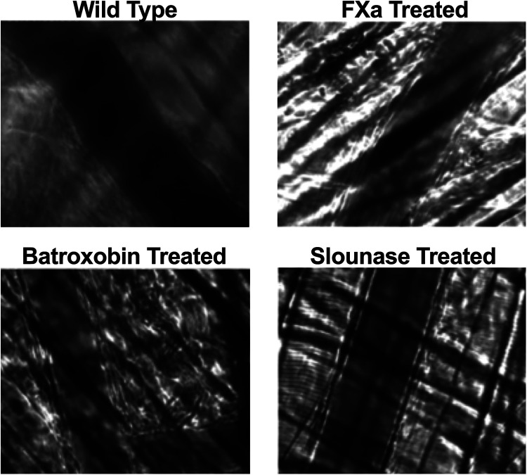
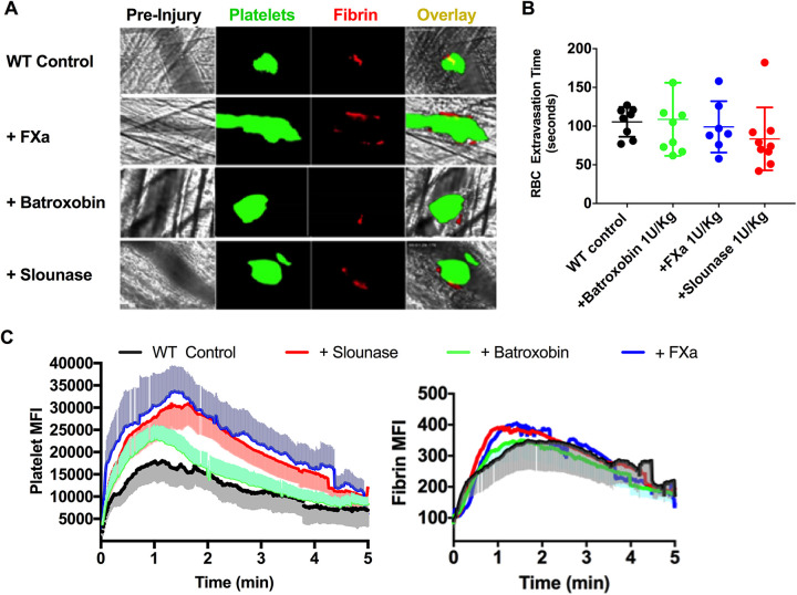
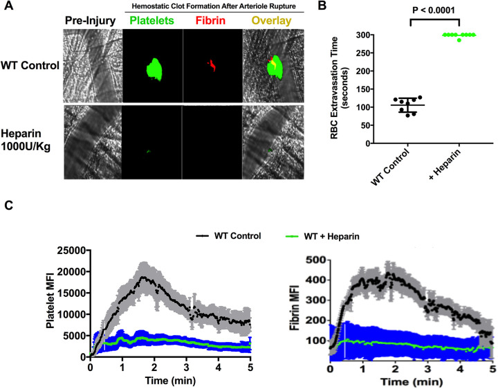
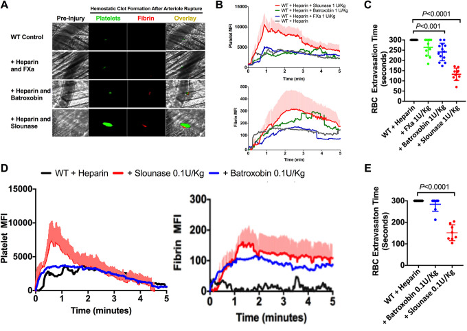
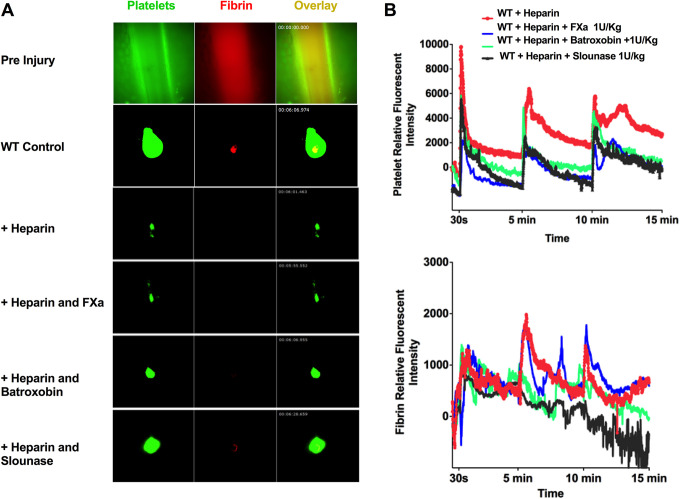
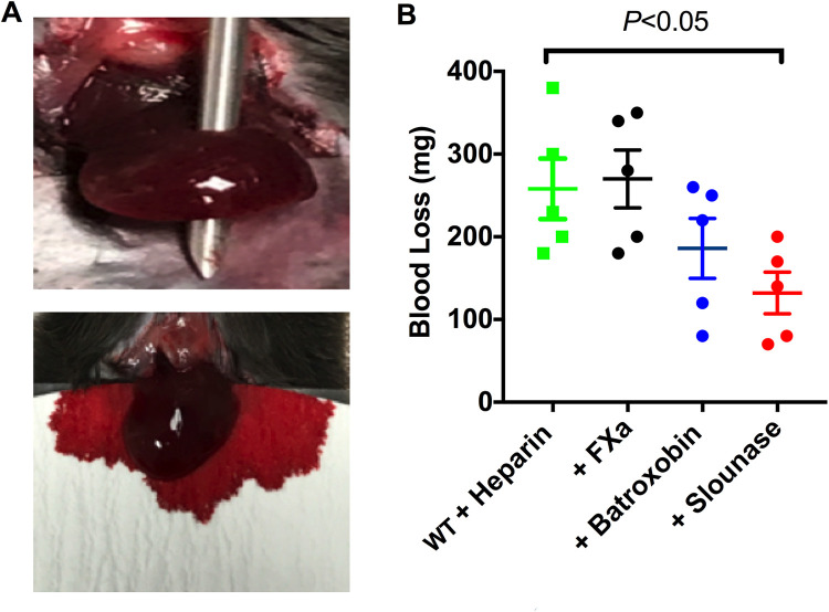
Uncontrolled bleeding associated with trauma and surgery is the leading cause of preventable death. Batroxobin, a snake venom-derived thrombin-like serine protease, has been shown to clot fibrinogen by cleaving fibrinopeptide A in a manner distinctly different from thrombin, even in the presence of heparin. The biochemical properties of batroxobin and its effect on coagulation have been well characterized in vitro. However, the efficacy of batroxobin on hemostatic clot formation in vivo is not well studied due to the lack of reliable in vivo hemostasis models. Here, we studied the efficacy of batroxobin and slounase, a batroxobin containing activated factor X, on hemostatic clot composition and bleeding using intravital microcopy laser ablation hemostasis models in micro and macro vessels and liver puncture hemostasis models in normal and heparin-induced hypocoagulant mice. We found that prophylactic treatment in wild-type mice with batroxobin, slounase and activated factor X significantly enhanced platelet-rich fibrin clot formation following vascular injury. In heparin-treated mice, batroxobin treatment resulted in detectable fibrin formation and a modest increase in hemostatic clot size, while activated factor X had no effect. In contrast, slounase treatment significantly enhanced both platelet recruitment and fibrin formation, forming a stable clot and shortening bleeding time and blood loss in wild-type and heparin-treated hypocoagulant mice. Our data demonstrate that, while batroxobin enhances fibrin formation, slounase was able to enhance hemostasis in normal mice and restore hemostasis in hypocoagulant conditions via the enhancement of fibrin formation and platelet activation, indicating that slounase is more effective in controlling hemorrhage.

Keywords: batroxobin; bleeding; coagulation; fibrin; hemostasis; platelet.


Conflict of interest statement

Declaration of Conflicting Interests: The author(s) declared the following potential conflicts of interest with respect to the research, authorship, and/or publication of this article: This work was supported by Lee’s Pharmaceutical Holdings limited. Dai is an employee of Zhaoke Pharmaceutical Co. Ltd. M. Li and B. X. Li is employer of Lee’s Pharmaceutical Holdings limited, the company which holds the patent for slounase.

Figures

Figure 1.
Figure 1.
Intravenous administration of slounase, batroxobin or activated factor X did not cause detectable platelet adhesion or platelet aggregate formation in the absence of vascular injury in vivo. Blood flow in cremaster microcirculation of WT mice was monitored in real-time and recorded under the intravital microscope. Circulating platelets were fluorescently labeled as green and detection of fibrin was achieved by injecting fluorescently conjugated anti-fibrin antibody as red in vivo. 1 U/kg of activated factor X, batroxobin, slounase, or control buffer were intravenously injected while real-time recording and continuously monitored for an extended period of time up to 90 minutes without inducing vascular injury. No platelet adhesion, aggregation or fibrin formation was detected in cremaster microcirculation prior to, during, or up to 90 minutes post injection. Representative pictures of overlaid bright field with fluorescent channels after subtracting fluorescent backgrounds (10 min post injection) show no fluorescent platelet adherence, aggregation, or fibrin formation on the cremaster arterioles vessel wall in the absence of injury in vivo following the injection of slounase (bottom right), batroxobin (bottom left), or activated factor X (top right) respectively.
Figure 2.
Figure 2.
Hemostatic clot formation was enhanced in WT normal coagulant mice pretreated with activated factor X, batroxobin or slounase. WT mice were pretreated with 1 U/kg of slounase, batroxobin, activated factor X or control buffer respectively and hemostatic response and bleeding were assessed by a laser-ablation puncture to the cremaster arterioles as described. (A) Representative images of hemostatic clot formation in response to laser-induced cremaster arteriole wall rupture. Platelet accumulation is shown in green, fibrin formation is shown in red and composite images of hemostatic clot formation are shown in yellow. (B) The time required for the cessation of RBC extravasation from arterioles in WT control mice and WT mice treated with 1U/kg of slounase, batroxobin, or activated factor X (Data from 2-3 independent injuries per mouse, 3 mice in each group. P < 0.001). (C) Dynamics of mean fluorescent intensity of platelets (left) and fibrin (right) in a hemostatic clot plotted as a function of time. WT mice were pretreated with slounase, batroxobin, activated factor X or control buffer and fluorescent intensity was recorded over 5 minutes. The shaded regions are representative of the standard error (SEM).
Figure 3.
Figure 3.
Heparin treatment in mice potently inhibited platelet accumulation and fibrin formation at the site of vascular injury in vivo and impaired hemostasis, resulting in prolonged bleeding in the laser-ablation cremaster arteriole puncture hemostasis model. WT mice were intravenously injected with saline (control) or 1000 U/kg heparin to inhibit coagulation in vivo then the hemostatic response and bleeding were assessed by laser-ablation puncture to the cremaster arterioles as described. The cremaster muscle arteriole wall was exposed to a high-intensity laser pulse to puncture a hole, and bleeding was monitored by RBC extravasation from the cremaster arterioles wall. The subsequent formation of a platelet-fibrin hemostatic clot was recorded in real-time under intravital microscopy. (A) Representative images of hemostatic clot formation in WT control mice (upper panel) and WT mice pretreated with heparin in response to laser-induced cremaster arteriole wall rupture. Platelet accumulation is shown in green, fibrin formation is shown in red and composite images are shown in yellow. (B) The time required for the cessation of RBC extravasation from arterioles in WT control mice (black) and WT mice treated with heparin (green) was analyzed by reviewing sequential images offline. (Data from 2 independent injuries per mouse, 4 mice in each group. P < 0.0001). (C) Dynamics of platelet accumulation and fibrin formation in response to injury were assesses by the changes in the fluorescent intensity of platelets (left) and fibrin (right) in a hemostatic clot in WT control mice (black) and WT mice treated with heparin (green). The shaded regions are representative of the standard error (SEM).
Figure 4.
Figure 4.
Slounase treatment restored hemostatic clot formation and shortened arterial bleeding time in heparin-induced hypocoagulant mice. WT mice were intravenously injected with 1000U/kg of heparin to induce a hypocoagulant condition. After confirming the absence of hemostatic response to injury using a laser-ablation puncture to the cremaster arteriole, mice were given intravenous treatment with 0.1 or 1 U/kg of activated factor X, batroxobin or slounase. Then, the hemostatic response to injury was continuously evaluated through RBC extravasation and hemostatic clot formation in real time under a microscope in response to the laser-ablation cremaster arteriole puncture as described. (A) Representative images of hemostatic clot formation in heparinized WT mice treated with control buffer or 1U/kg of activated factor X, batroxobin, or slounase, respectively. (B) Dynamics of fluorescent intensity of platelets (top) and fibrin (bottom) in a hemostatic clot in heparinized WT mice treated with saline or 1U/kg of activated factor X, batroxobin or slounase (P < 0.001). The shaded regions are representative of the standard error (SEM). (C) The time required for the cessation of RBC extravasation from arterioles in heparinized WT mice treated with saline or 1 U/kg (left) of activated factor X, batroxobin or slounase. (Data from 2 independent injuries per mouse, 3 mice in each group. P<0.0001). (D) Dynamics of fluorescent intensity of platelets (left) and fibrin (right) in a hemostatic clot in heparinized WT mice treated with saline or 0.1 U/kg of activated factor X, batroxobin or slounase. The shaded regions are representative of the standard error (SEM). (E) The time required for the cessation of RBC extravasation from arterioles in heparinized WT mice treated with saline or 0.1 U/kg of activated factor X, batroxobin or slounase. (Data from 2 independent injuries per mouse, 3 mice in each group. P < 0.0001).
Figure 5.
Figure 5.
Slounase treatment enhances hemostatic plug formation in the saphenous vein of hypocoagulant mice. WT mice were intravenously injected with control buffer or 1000 U/kg of heparin to induce a hypocoagulant condition. Subgroups of heparin-treated mice were also were given intravenous treatment with 1 U/kg of activated factor X, batroxobin and slounase. Hemostatic plug formation in the saphenous vein wall was assessed using laser-ablation cremaster arteriole hemostasis model under intravital microscopy as described. (A) Representative images of the saphenous vein in multi-channel prior to vascular injury (with a fluorescent background) are shown in the top panel. Representative images of hemostatic clot formation after vascular injury (2nd laser injury after the subtraction of the fluorescent background) in control WT mice, WT mice treated with heparin alone, or further treated with 1 U/kg of activated factor X, batroxobin or slounase, are shown below as indicated. Within the hemostatic plug that formed at the site of injury on the saphenous vessel wall, platelet accumulation is shown in green, fibrin formation is shown in red and composite images are shown in yellow. (B) Quantitative analysis of platelet accumulation and fibrin formation in the hemostatic clot with repetitive vascular injury to the saphenous vein. Times of vascular injury are indicated at 30 seconds and repeated at 5 and 10 minutes. The kinetic curves represent the relative platelet (top) and fibrin (bottom) fluorescent intensity (n = 6; 2 independent injuries in each mouse, 3 mice in each group). Slounase treatment in heparinized WT mice partially restored clot formation by enhancing platelet adhesion and accumulation as well as fibrin formation (P < 0.05) in the saphenous vein when compared with batroxobin and activated factor X treatment in heparin-treated hypocoagulant mice.
Figure 6.
Figure 6.
Slounase treatment reduces total blood loss in the liver wound model. (A) Mice were treated with 1000U/kg of heparin followed by an intravenous injection with 1U/kg of slounase, batroxobin, or activated factor X respectively. Blood loss in mice was assessed using a pre-weighed filter paper following a needle injury to the left lobe of the mouse liver as described (top). Bleeding in hypocoagulant mice induced by heparin following the injury is shown (bottom). (B) Total amount of blood lost in mice treated with heparin alone or further treated with activated factor X, batroxobin or slounase. Slounase treatment effectively reduced the blood loss when compared to control buffer treatment in the hypocoagulant condition induced by heparin treatment (n = 5 mice /group, P < 0.05).

Similar articles

Cited by

  • Smart thrombosis inhibitors without bleeding side effects via charge tunable ligand design.
    La CC, Smith SA, Vappala S, Adili R, Luke CE, Abbina S, Luo HD, Chafeeva I, Drayton M, Creagh LA, de Guadalupe Jaraquemada-Peláez M, Rhoads N, Kalathottukaren MT, Henke PK, Straus SK, Du C, Conway EM, Holinstat M, Haynes CA, Morrissey JH, Kizhakkedathu JN. La CC, et al. Nat Commun. 2023 Apr 26;14(1):2177. doi: 10.1038/s41467-023-37709-0. Nat Commun. 2023. PMID: 37100783 Free PMC article.

References

    1. Rossaint R, Bouillon B, Cerny V, et al. The European guideline on management of major bleeding and coagulopathy following trauma: fourth edition. Crit Care. 2016;20:100. - PMC - PubMed
    1. Kauvar DS, Lefering R, Wade CE. Impact of hemorrhage on trauma outcome: an overview of epidemiology, clinical presentations, and therapeutic considerations. J Trauma. 2006;60(6 Suppl):S3–11. - PubMed
    1. Ruggeri ZM. Platelets in atherothrombosis. Nat Med. 2002;8(11):1227–1234. - PubMed
    1. Ni H, Freedman J. Platelets in hemostasis and thrombosis: role of integrins and their ligands. Transfus Apher Sci. 2003;28(3):257–264. - PubMed
    1. Jackson SP. The growing complexity of platelet aggregation. Blood. 2007;109(12):5087–5095. - PubMed

LinkOut - more resources