Review
. 2021 May 28;135(10):1311-1332.
doi: 10.1042/CS20200301.

Molecular mechanisms of anthracycline cardiovascular toxicity

Affiliations
Review

Molecular mechanisms of anthracycline cardiovascular toxicity

Anna Narezkina et al. Clin Sci (Lond). .

Abstract

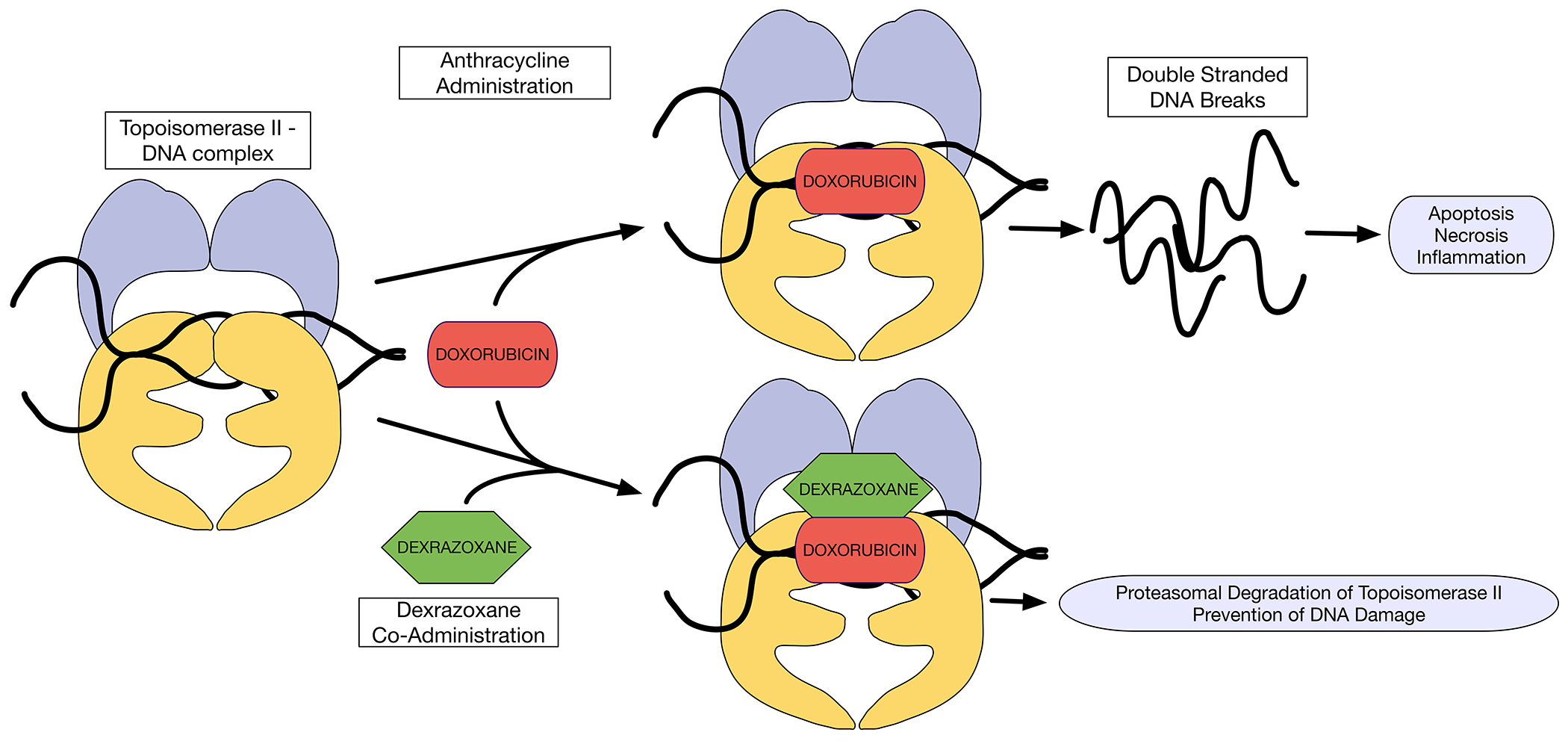
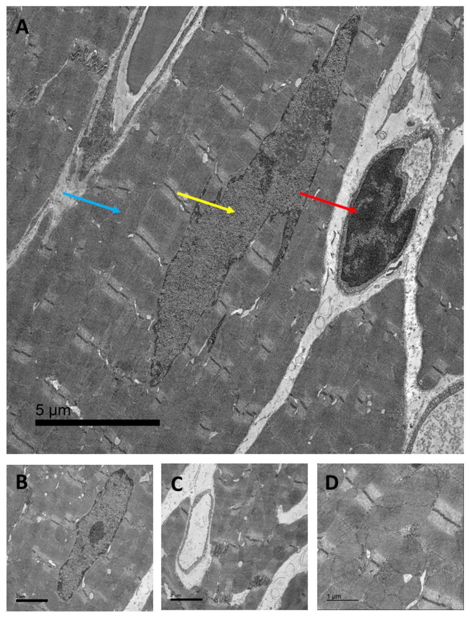
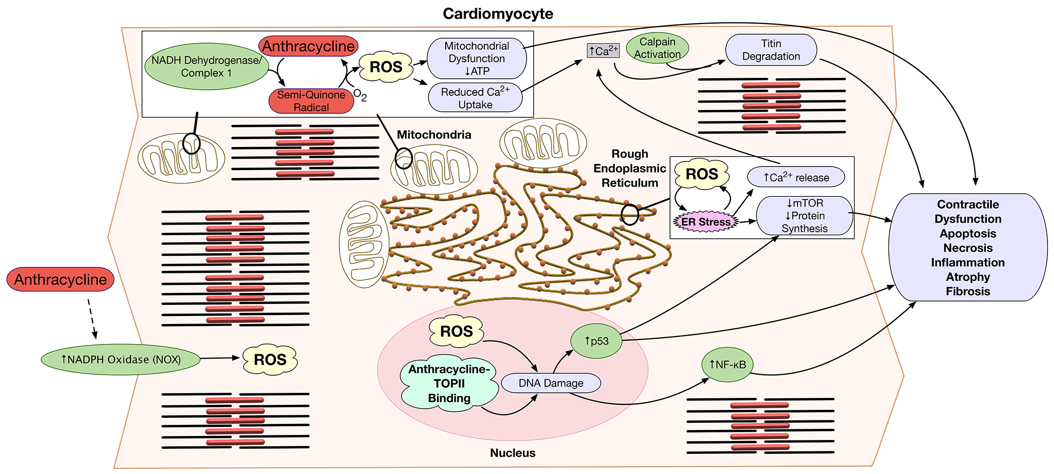
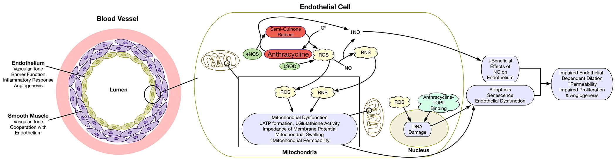
Anthracyclines are effective chemotherapeutic agents, commonly used in the treatment of a variety of hematologic malignancies and solid tumors. However, their use is associated with a significant risk of cardiovascular toxicities and may result in cardiomyopathy and heart failure. Cardiomyocyte toxicity occurs via multiple molecular mechanisms, including topoisomerase II-mediated DNA double-strand breaks and reactive oxygen species (ROS) formation via effects on the mitochondrial electron transport chain, NADPH oxidases (NOXs), and nitric oxide synthases (NOSs). Excess ROS may cause mitochondrial dysfunction, endoplasmic reticulum stress, calcium release, and DNA damage, which may result in cardiomyocyte dysfunction or cell death. These pathophysiologic mechanisms cause tissue-level manifestations, including characteristic histopathologic changes (myocyte vacuolization, myofibrillar loss, and cell death), atrophy and fibrosis, and organ-level manifestations including cardiac contractile dysfunction and vascular dysfunction. In addition, these mechanisms are relevant to current and emerging strategies to diagnose, prevent, and treat anthracycline-induced cardiomyopathy. This review details the established and emerging data regarding the molecular mechanisms of anthracycline-induced cardiovascular toxicity.

Keywords: anthracycline; cardiomyopathy; cardiotoxicity; reactive oxygen species; topoisomerases.


Conflict of interest statement

Dr. Zemljic-Harpf receives funding from the Merck Investigator Studies Program and Scientific Engagements.

Figures

Figure 1.
Figure 1.. Anthracycline effect on Topoisomerase 2β, and its inhibition by dexrazoxane.
Doxorubicin intercalates DNA and inhibits Top2β. Doxorubicin forms cleavable complexes with DNA-bound Top2β, which may lead to DNA breaks and induce cell death. Dexrazoxane binds to DNA-bound Top2β, and triggers proteasomal degradation of Top2β, thereby reducing formation of doxorubicin-poisoned Top2β cleavage complexes. DNA = deoxyribonucleic acid; Top2β = Topoisomerase 2β
Figure 2.
Figure 2.. Myocardial cells and sub-cellular compartments affected by anthracycline-induced topoisomerase II inhibition.
Transmission electron microscopic scan of murine cardiac tissue: A) Arrows indicate cardiac mitochondria (blue arrow, mitochondrial DNA), the cardiac nucleus (yellow arrow, nuclear DNA), and cardiac endothelial cells (red arrow, endothelial cell DNA, 2,500 x, scale bar = 5 μm). B + C) cardiomyocyte nucleus and cardiac capillary formed by a single endothelial cell, respectively (5,000x, scale bar = 2 μm), and D) cardiac interfibrillar mitochondria (10,000x, scale bar = 1 μm). DNA = deoxyribonucleic acid
Figure 3.
Figure 3.. Molecular pathways affected by anthracyclines in cardiac myocytes.
In myocytes anthracyclines induce ROS generation by mitochondria and NADPH oxidase (NOX), reduce mitochondrial respiration, induce ER-stress, alter Ca2+ signaling, and induce DNA damage. NADH dehydrogenase of complex I of the mitochondrial electron transport chain reduces the quinone moiety of anthracyclines to a semiquinone radical. In the presence of molecular oxygen, the semiquinone auto-oxidizes to generate the parent anthracycline and a superoxide anion. The effects of anthracyclines on the electron transport chain and increased ROS generation cause mitochondrial dysfunction, decreased ATP synthesis, and reduced Ca++ uptake. Increased ROS production induces ER-stress, further increasing cytoplasmic Ca++ concentrations due to ER Ca++ leak. ER stress also inhibits mTOR survival signaling and thereby reduces protein synthesis. ER-stress and mTOR inhibition trigger inflammation, and cell death signaling. In addition, anthracyclines induce activation of the NOX transmembrane enzyme complex, further increasing production of ROS. Anthracycline binding to TopII in the nucleus, and the direct effect of ROS may cause irreversible DNA damage. Anthracycline-induced DNA damage and oxidative stress lead to p53 tumor suppressor activation, that modulates mTOR pathway. ROS and DNA damage result in NF-kB activation. These molecular pathways lead to contractile dysfunction, cardiac atrophy, fibrosis, apoptosis, necrosis, and inflammation. ATP = adenosine triphosphate; DNA = deoxyribonucleic acid; ER = endoplasmic reticulum; ROS = reactive oxygen species; TOPII = topoisomerase II
Figure 4.
Figure 4.. Effects of anthracyclines on the vascular endothelium.
Within the vascular endothelium, anthracyclines result in the generation of reactive oxygen species (ROS) primarily via endothelial nitric oxide synthase (eNOS). Interactions of ROS with nitric oxide (NO) results in the formation of peroxynitrites (reactive nitrogen species; RNS) and result in reductions in NO. These reactive species result in mitochondrial dysfunction. DNA damage may result from ROS and topoisomerase II-mediated double stranded DNA breaks. Through these mechanisms, anthracyclines may cause endothelial apoptosis, senescence, and dysfunction resulting in impaired endothelial dependent vascular dilation, increased permeability, and impaired angiogenesis. ATP = adenosine triphosphate; DNA = deoxyribonucleic acid; eNOS = endothelial nitric oxide synthase; RNS = reactive nitrogen species; ROS = reactive oxygen species; SOD = superoxide dismutase; TOPII = topoisomerase II

Similar articles

Cited by

References

    1. McGowan JV, Chung R, Maulik A, Piotrowska I, Walker JM and Yellon DM (2017) Anthracycline Chemotherapy and Cardiotoxicity. Cardiovasc. Drugs Ther 31, 63–75. - PMC - PubMed
    1. Mulrooney DA, Hyun G, Ness KK, Ehrhardt MJ, Yasui Y, Duprez D, Howell RM, Leisenring WM, Constine LS, Tonorezos E, et al. (2020) Major cardiac events for adult survivors of childhood cancer diagnosed between 1970 and 1999: report from the Childhood Cancer Survivor Study cohort. The BMJ 368. - PMC - PubMed
    1. https://www.nccn.org/professionals/physician_gls/pdf/sarcoma.pdf.
    1. Giordano SH, Lin Y-L, Kuo YF, Hortobagyi GN and Goodwin JS (2012) Decline in the Use of Anthracyclines for Breast Cancer. J. Clin. Oncol 30, 2232–2239. - PMC - PubMed
    1. Armstrong GT, Liu Q, Yasui Y, Neglia JP, Leisenring W, Robison LL and Mertens AC (2009) Late Mortality Among 5-Year Survivors of Childhood Cancer: A Summary From the Childhood Cancer Survivor Study. J. Clin. Oncol 27, 2328–2338. - PMC - PubMed

Publication types

MeSH terms