

Rare Neurovascular Diseases in Korea: Classification and Related Genetic Variants
- PMID: 34047503
- PMCID: PMC8316781
- DOI: 10.3348/kjr.2020.1171
Rare Neurovascular Diseases in Korea: Classification and Related Genetic Variants
Abstract
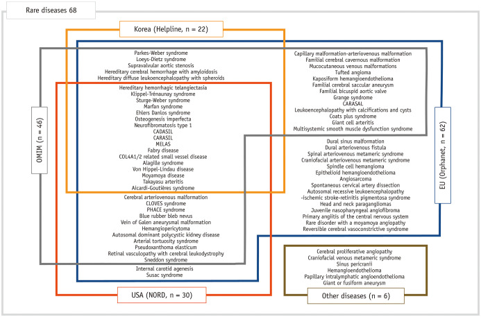
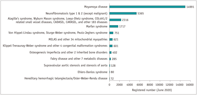
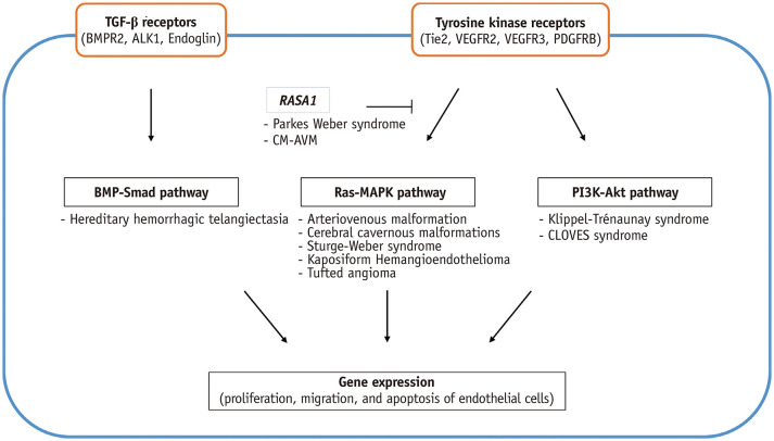
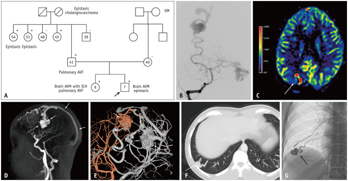
Rare neurovascular diseases (RNVDs) have not been well-recognized in Korea. They involve the central nervous system and greatly affect the patients' lives. However, these diseases are difficult to diagnose and treat due to their rarity and incurability. We established a list of RNVDs by referring to the previous literature and databases worldwide to better understand the diseases and their current management status. We categorized 68 RNVDs based on their pathophysiology and clinical manifestations and estimated the prevalence of each disease in Korea. Recent advances in genetic, molecular, and developmental research have enabled further understanding of these RNVDs. Herein, we review each disease, while considering its classification based on updated pathologic mechanisms, and discuss the management status of RNVD in Korea.
Keywords: Classification; Diagnosis; Genetics; Neurovascular; Rare diseases.
Copyright © 2021 The Korean Society of Radiology.
Conflict of interest statement
The authors have no potential conflicts of interest to disclose.
Figures
Similar articles
-
Prevalence of systemic lupus erythematosus in South Korea: an administrative database study.J Epidemiol. 2014;24(4):295-303. doi: 10.2188/jea.je20120204. Epub 2014 May 24. J Epidemiol. 2014. PMID: 24857955 Free PMC article.
-
The cumulative incidence and trends of rare diseases in South Korea: a nationwide study of the administrative data from the National Health Insurance Service database from 2011-2015.Orphanet J Rare Dis. 2019 Feb 18;14(1):49. doi: 10.1186/s13023-019-1032-6. Orphanet J Rare Dis. 2019. PMID: 30777110 Free PMC article.
-
Prevalence and incidence of sarcoidosis in Korea: a nationwide population-based study.Respir Res. 2018 Aug 28;19(1):158. doi: 10.1186/s12931-018-0871-3. Respir Res. 2018. PMID: 30153830 Free PMC article.
-
Prevalence, burden, and clinical management of migraine in China, Japan, and South Korea: a comprehensive review of the literature.J Headache Pain. 2019 Dec 5;20(1):111. doi: 10.1186/s10194-019-1062-4. J Headache Pain. 2019. PMID: 31805851 Free PMC article. Review.
-
Current status of functional dyspepsia in Korea.Korean J Intern Med. 2014 Mar;29(2):156-65. doi: 10.3904/kjim.2014.29.2.156. Epub 2014 Feb 27. Korean J Intern Med. 2014. PMID: 24648796 Free PMC article. Review.
Cited by
-
Clinical Manifestation and Radiologic Patterns of Spontaneous Cervicocephalic Dissection According to the Anatomic Location: A Single-Center Analysis in Korean Patients.Neurointervention. 2022 Jul;17(2):78-86. doi: 10.5469/neuroint.2022.00143. Epub 2022 Jun 13. Neurointervention. 2022. PMID: 35692184 Free PMC article.
-
Flow Diverter Treatment Using a Flow Re-Direction Endoluminal Device for Unruptured Intracranial Vertebral Artery Dissecting Aneurysm: Single-Center Case Series and Technical Considerations.Neurointervention. 2023 Jul;18(2):114-122. doi: 10.5469/neuroint.2023.00199. Epub 2023 Jun 20. Neurointervention. 2023. PMID: 37337396 Free PMC article.
-
The role of public health in rare diseases: hemophilia as an example.Front Public Health. 2025 Mar 20;13:1450625. doi: 10.3389/fpubh.2025.1450625. eCollection 2025. Front Public Health. 2025. PMID: 40182514 Free PMC article. Review.
-
Physiologic Flow Diversion Coiling Technique for Wide-Necked Aneurysms with an Asymmetric Bidirectional Flow at the Aneurysm Neck.Neurointervention. 2022 Nov;17(3):133-142. doi: 10.5469/neuroint.2022.00311. Epub 2022 Sep 28. Neurointervention. 2022. PMID: 36167347 Free PMC article.
-
Effect of bisphenol A on the neurological system: a review update.Arch Toxicol. 2024 Jan;98(1):1-73. doi: 10.1007/s00204-023-03614-0. Epub 2023 Oct 19. Arch Toxicol. 2024. PMID: 37855918 Free PMC article. Review.
References
-
- Richter T, Nestler-Parr S, Babela R, Khan ZM, Tesoro T, Molsen E, et al. Rare disease terminology and definitions—a systematic global review: report of the ISPOR rare disease special interest group. Value Health. 2015;18:906–914. - PubMed
-
- Vanaman MJ, Hervey-Jumper SL, Maher CO. Pediatric and inherited neurovascular diseases. Neurosurg Clin N Am. 2010;21:427–441. - PubMed
Publication types
MeSH terms
LinkOut - more resources
Full Text Sources
Other Literature Sources
