Review
. 2021 Aug;22(8):1310-1322.
doi: 10.3348/kjr.2020.1299. Epub 2021 May 20.

Immunotherapy-Related Imaging Findings in Patients with Gynecological Malignancies: What Radiologists Need to Know

Affiliations
Review

Immunotherapy-Related Imaging Findings in Patients with Gynecological Malignancies: What Radiologists Need to Know

Luca Russo et al. Korean J Radiol. 2021 Aug.

Abstract

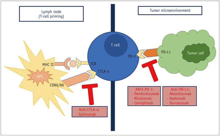
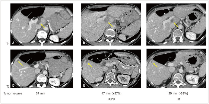
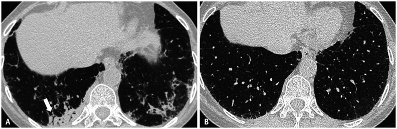
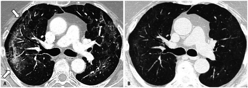
Immunotherapy is an effective treatment option for gynecological malignancies. Radiologists dealing with gynecological patients undergoing treatment with immune checkpoint inhibitors should be aware of unconventional immune-related imaging features for the evaluation of tumor response and immune-related adverse events. In this paper, immune checkpoint inhibitors used for gynecological malignancies and their mechanisms of action are briefly presented. In the second part, patterns of pseudoprogression are illustrated, and different forms of immune-related adverse events are discussed.

Keywords: Adverse drug event; Checkpoint inhibitors; Gynaecological oncology; Pseudoprogression.


Conflict of interest statement

The authors have no potential conflicts of interest to disclose.

Figures

Fig. 1
Fig. 1. Mechanisms of action of CTLA-4 and PD-1.
CTLA-4 binds CD80/86 expressed on antigen-presenting cell, inhibiting T cell. PD-1 binds PD-L1, expressed on tumor cell, resulting in suppression of immune response. APC = antigen-presenting cell, CD = cluster of differentiation, CTLA-4 = cytotoxic T-limphocyte antigen 4, MHC = major histocompatibility complex, PD-1 = programmed cell death protein 1, PD-L1 = programmed cell death protein 1 ligand, TCR = T-cell receptor
Fig. 2
Fig. 2. iRECIST criteria for evaluation of a 51-year-old female with ovarian cancer treated with pembrolizumab (anti PD-1).
A, D. Baseline CT shows two peritoneal subglissonian metastases (arrows; T1: 17 mm; T2: 10 mm). B, E. On a 3-month follow-up, both lesions enlarged with an increase of 27% of the target lesions leading to an iUPD according to iRECIST. C, F. After 8 weeks, both lesions decreased in size, compared to baseline examination (−33%). iRECIST = included in the Response Evaluation Criteria in Solid Tumors, iUPD = unconfirmed progressive disease, PD-1 = programmed cell death protein 1, PR = partial response
Fig. 3
Fig. 3. Most common immune-related adverse events in female.
Fig. 4
Fig. 4. Pneumonitis with an organizing pneumonia pattern.
57-year-old female with cervival cancer in treatment with atezolizumab showing mild dyspnea. A. Follow-up CT scan performed 5 weeks after atezolizumab initiation shows bilateral lower lobes consolidative opacities. B. CT scan performed 4 weeks after atezolizumab interruption and corticosteroids administration shows pneumonitis resolution with residual subpleural reticular opacities.
Fig. 5
Fig. 5. Pneumonitis with a NSIP pattern.
72 year-old female with ovarian cancer in treatment with durvalumab showing no significant respiratory symptoms A. Follow-up CT scan performed 6 weeks after durvalumab initiation shows bilateral confluent ground-glass and reticular opacities with subpleural sparing (arrows). B. CT scan performed 4 weeks after durvalumab interruption and corticosteroids administration shows partial resolution of NSIP. NSIP = non-specific interstitial pneumonia
Fig. 6
Fig. 6. Colitis.
58-year-old female with ovarian cancer presenting at emergency department with abdominal pain and diarrhea, 10 weeks after Pembrolizumab initiation. A, B. Coronal (A) and axial (B) contrast-enhanced abdominal CT show wall thickening of the descending colon (arrows) and surrounding fat stranding (arrowheads). C, D. Coronal (C) and axial (D) contrast-enhanced CT image, acquired 6 weeks after pembrolizumab interruption and corticosteroids administration shows normal appearance of descending colon (arrows).
Fig. 7
Fig. 7. Enteritis.
73-year-old female with endometrial cancer in treatment performing follow-up CT and pelvis MRI. A. Follow-up CT scan performed 15 weeks after Pembrolizumab initiation shows wall thickening of an ileal loop (arrow). B. Axial fast spin echo T2-weighted images shows wall thickening and mild hyperintensity of submucosal layer (arrow), representing oedema. C. Axial T1-weighted fat-sat post-gadolinium images better show stratified pattern of the ileal wall thickening with mucosal enhancement (arrow) and low-intensity submucosal oedema.
Fig. 8
Fig. 8. Pancreatitis.
66-year-old female with cervix cancer presenting at emergency department with right abdominal pain, 10 weeks after Atezolizumab initiation. A. Contrast-enhanced CT scan shows pancreatic head enlargement associated to focal hypo-enhancement (arrow). B. Axial T1-weighted fat-sat images clearly show focal hypointensity on pancreatic head (arrow). C. Diffusion-weighted images shows significant diffusion restriction in the pancreatic head. D. CT scan performed 6 weeks after treatment interruption and corticosteroids administration shows normal appearence of the pancreas.
Fig. 9
Fig. 9. Cholecystitis.
71-year-old female with ovarian cancer presenting at emergency department with right upper abdominal pain, 12 weeks after pembrolizumab initiation. Coronal-reconstructed abdominal CT scan shows mucosal enhancement (arrow) and pericholecistic fluid collection (arrowhead).

Similar articles

Cited by

References

    1. Bhatla N, Aoki D, Sharma DN, Sankaranarayanan R. Cancer of the cervix uteri. Int J Gynaecol Obstet. 2018;143 Suppl 2:22–36. - PubMed
    1. Ferlay J, Soerjomataram I, Dikshit R, Eser S, Mathers C, Rebelo M, et al. Cancer incidence and mortality worldwide: sources, methods and major patterns in GLOBOCAN 2012. Int J Cancer. 2015;136:E359–E386. - PubMed
    1. Mihor A, Tomsic S, Zagar T, Lokar K, Zadnik V. Socioeconomic inequalities in cancer incidence in Europe: a comprehensive review of population-based epidemiological studies. Radiol Oncol. 2020;54:1–13. - PMC - PubMed
    1. Siegel RL, Miller KD, Jemal A. Cancer statistics, 2018. CA Cancer J Clin. 2018;68:7–30. - PubMed
    1. Hodi FS, O'Day SJ, McDermott DF, Weber RW, Sosman JA, Haanen JB, et al. Improved survival with ipilimumab in patients with metastatic melanoma. N Engl J Med. 2010;363:711–723. - PMC - PubMed

Substances