Establishing functional lentiviral vector production in a stirred bioreactor for CAR-T cell therapy
- PMID: 34047682
- PMCID: PMC8806440
- DOI: 10.1080/21655979.2021.1931644
Establishing functional lentiviral vector production in a stirred bioreactor for CAR-T cell therapy
Abstract
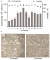
As gene delivery tools, lentiviral vectors (LV) have broad applications in chimeric antigen receptor therapy (CAR-T). Large-scale production of functional LV is limited by the adherent, serum-dependent nature of HEK293T cells used in the manufacturing. HEK293T adherent cells were adapted to suspension cells in a serum-free medium to establish large-scale processes for functional LV production in a stirred bioreactor without micro-carriers. The results showed that 293 T suspension was successfully cultivated in F media (293 CD05 medium and SMM293-TII with 1:1 volume ratio), and the cells retained the capacity for LV production. After cultivation in a 5.5 L bioreactor for 4 days, the cells produced 1.5 ± 0.3 × 10⁷ TU/mL raw LV, and the lentiviral transduction efficiency was 48.6 ± 2.8% in T Cells. The yield of LV equaled to the previous shake flask. The critical process steps were completed to enable a large-scale LV production process. Besides, a cryopreservation solution was developed to reduce protein involvement, avoid cell grafting and reduce process cost. The process is cost-effective and easy to scale up production, which is expected to be highly competitive.
Keywords: CAR-T cell therapy; Functional; HEK293T cells; lentiviral vectors; stirred bioreactors.
Figures


Similar articles
-
Scalable Lentiviral Vector Production Using Stable HEK293SF Producer Cell Lines.Hum Gene Ther Methods. 2017 Dec;28(6):330-339. doi: 10.1089/hgtb.2017.086. Epub 2017 Nov 21. Hum Gene Ther Methods. 2017. PMID: 28826344 Free PMC article.
-
Generation of stable suspension producer cell lines for serum-free lentivirus production.Biotechnol J. 2024 May;19(5):e2400090. doi: 10.1002/biot.202400090. Biotechnol J. 2024. PMID: 38719592
-
Optimizing lentiviral vector formulation conditions for efficient ex vivo transduction of primary human T cells in chimeric antigen receptor T-cell manufacturing.Cytotherapy. 2024 Sep;26(9):1084-1094. doi: 10.1016/j.jcyt.2024.04.002. Epub 2024 Apr 7. Cytotherapy. 2024. PMID: 38661611
-
Lentiviral Vector Bioprocessing.Viruses. 2021 Feb 9;13(2):268. doi: 10.3390/v13020268. Viruses. 2021. PMID: 33572347 Free PMC article. Review.
-
Production of CAR T-cells by GMP-grade lentiviral vectors: latest advances and future prospects.Crit Rev Clin Lab Sci. 2019 Sep;56(6):393-419. doi: 10.1080/10408363.2019.1633512. Epub 2019 Jul 17. Crit Rev Clin Lab Sci. 2019. PMID: 31314617 Review.
Cited by
-
Genetic alteration of SJ293TS cells and modification of serum-free media enhances lentiviral vector production.Mol Ther Methods Clin Dev. 2024 May 21;32(2):101270. doi: 10.1016/j.omtm.2024.101270. eCollection 2024 Jun 13. Mol Ther Methods Clin Dev. 2024. PMID: 38883976 Free PMC article.
-
Recent Advances in the Development of Bioreactors for Manufacturing of Adoptive Cell Immunotherapies.Bioengineering (Basel). 2022 Dec 15;9(12):808. doi: 10.3390/bioengineering9120808. Bioengineering (Basel). 2022. PMID: 36551014 Free PMC article. Review.
References
-
- McCarron A, Donnelley M, McIntyre C, et al. Challenges of up-scaling lentivirus production and processing. J Biotechnol. 2016;240:23–30. - PubMed
Publication types
MeSH terms
LinkOut - more resources
Full Text Sources
Other Literature Sources
Research Materials
