. 2021 Jul 1;78(7):853-863.
doi: 10.1001/jamaneurol.2021.1509.

Clinicopathologic Features of Oculopharyngodistal Myopathy With LRP12 CGG Repeat Expansions Compared With Other Oculopharyngodistal Myopathy Subtypes

Theerawat Kumutpongpanich  1   2 Masashi Ogasawara  1   2 Ayami Ozaki  1   2 Hiroyuki Ishiura  3 Shoji Tsuji  3 Narihiro Minami  1   2 Shinichiro Hayashi  1   2 Satoru Noguchi  1   2 Aritoshi Iida  2 Ichizo Nishino  1   2 OPDM_LRP12 Study GroupMadoka Mori-Yoshimura  4 Yasushi Oya  4 Kenjiro Ono  5 Toshio Shimizu  6 Akihiro Kawata  6 Shun Shimohama  7 Keiko Toyooka  8 Kaoru Endo  9 Shuta Toru  10 Oga Sasaki  11 Kenji Isahaya  11 Masanori P Takahashi  12 Kazuo Iwasa  13 Jun-Ichi Kira  14 Tatsuya Yamamoto  15 Michi Kawamoto  16 Tadanori Hamano  17 Kazuma Sugie  18 Nobuyuki Eura  18 Tomo Shiota  18 Mizuho Koide  19 Kanako Sekiya  20 Hideaki Kishi  21 Takuto Hideyama  22 Shigeru Kawai  23 Satoshi Yanagimoto  23 Hiroyasu Sato  24 Hajime Arahata  25 Shigeo Murayama  26 Kayoko Saito  27 Hideo Hara  28 Takashi Kanda  29 Hiroshi Yaguchi  30 Noboru Imai  31 Yuichi Kawagashira  32 Mitsuru Sanada  33 Kazuki Obara  34 Misako Kaido  35 Minori Furuta  36 Takashi Kurashige  37 Wataru Hara  38 Daisuke Kuzume  39 Mamoru Yamamoto  40 Jun Tsugawa  41 Hitaru Kishida  42 Naoki Ishizuka  43 Kohei Morimoto  44 Yukio Tsuji  44 Atsuko Tsuneyama  45 Atsuhiro Matsuno  46 Ryo Sasaki  47 Daigo Tamakoshi  48 Erika Abe  49 Shinichiro Yamada  50 Akiyuki Uzawa  15
Affiliations

Clinicopathologic Features of Oculopharyngodistal Myopathy With LRP12 CGG Repeat Expansions Compared With Other Oculopharyngodistal Myopathy Subtypes

Theerawat Kumutpongpanich et al. JAMA Neurol. .

Abstract

Importance: Repeat expansion of CGG in LRP12 has been identified as the causative variation of oculopharyngodistal myopathy (OPDM). However, to our knowledge, the clinicopathologic features of OPDM with CGG repeat expansion in LRP12 (hereafter referred to as OPDM_LRP12) remain unknown.

Objective: To identify and characterize the clinicopathologic features of patients with OPDM_LRP12.

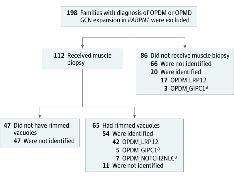
Design, setting, and participants: This case series included 208 patients with a clinical or clinicopathologic diagnosis of oculopharyngeal muscular dystrophy (OPDM) from January 1, 1978, to December 31, 2020. Patients with GCN repeat expansions in PABPN1 were excluded from the study. Repeat expansions of CGG in LRP12 were screened by repeat primed polymerase chain reaction and/or Southern blot.

Main outcomes and measures: Clinical information, muscle imaging data obtained by either computed tomography or magnetic resonance imaging, and muscle pathologic characteristics.

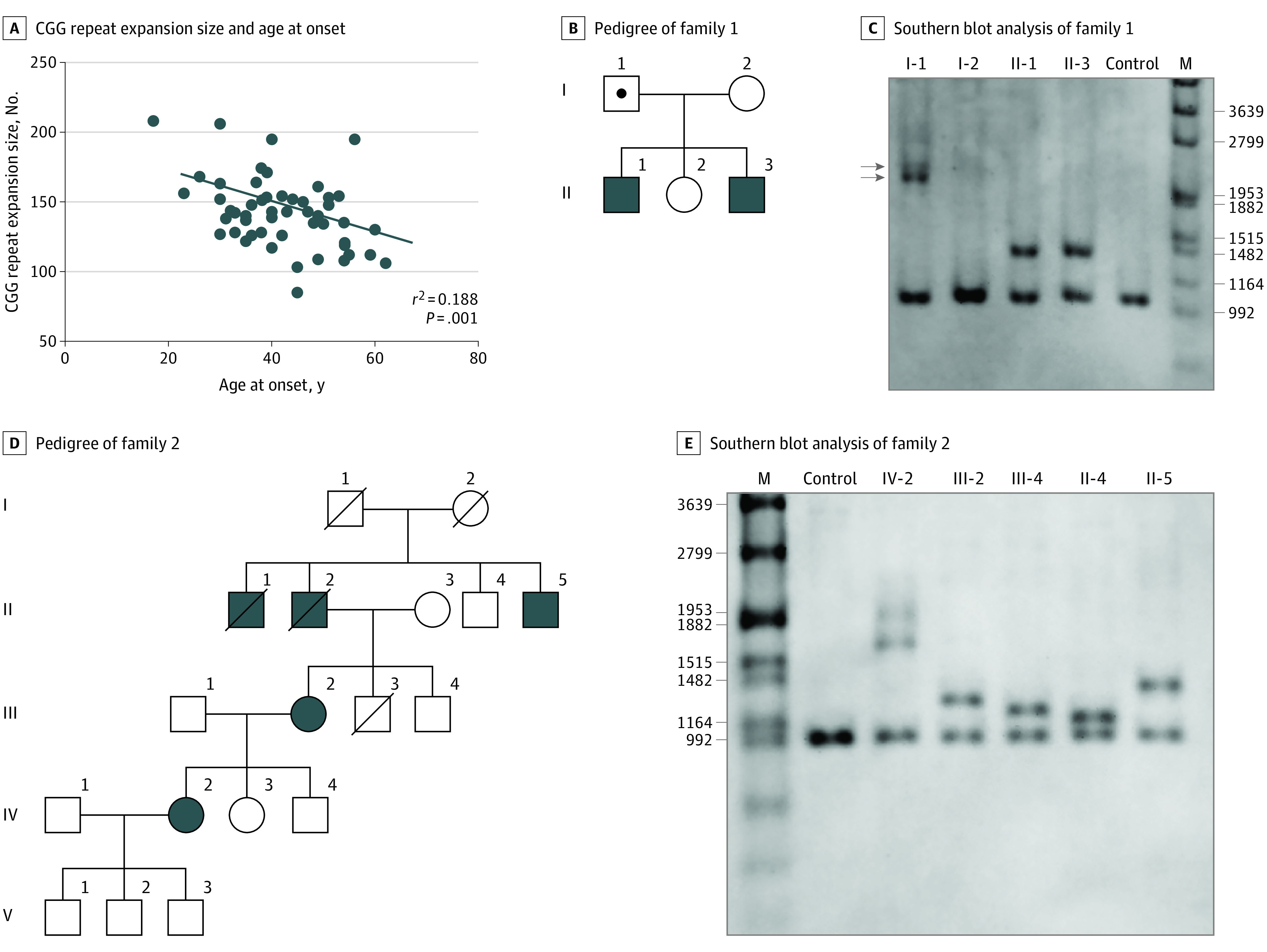
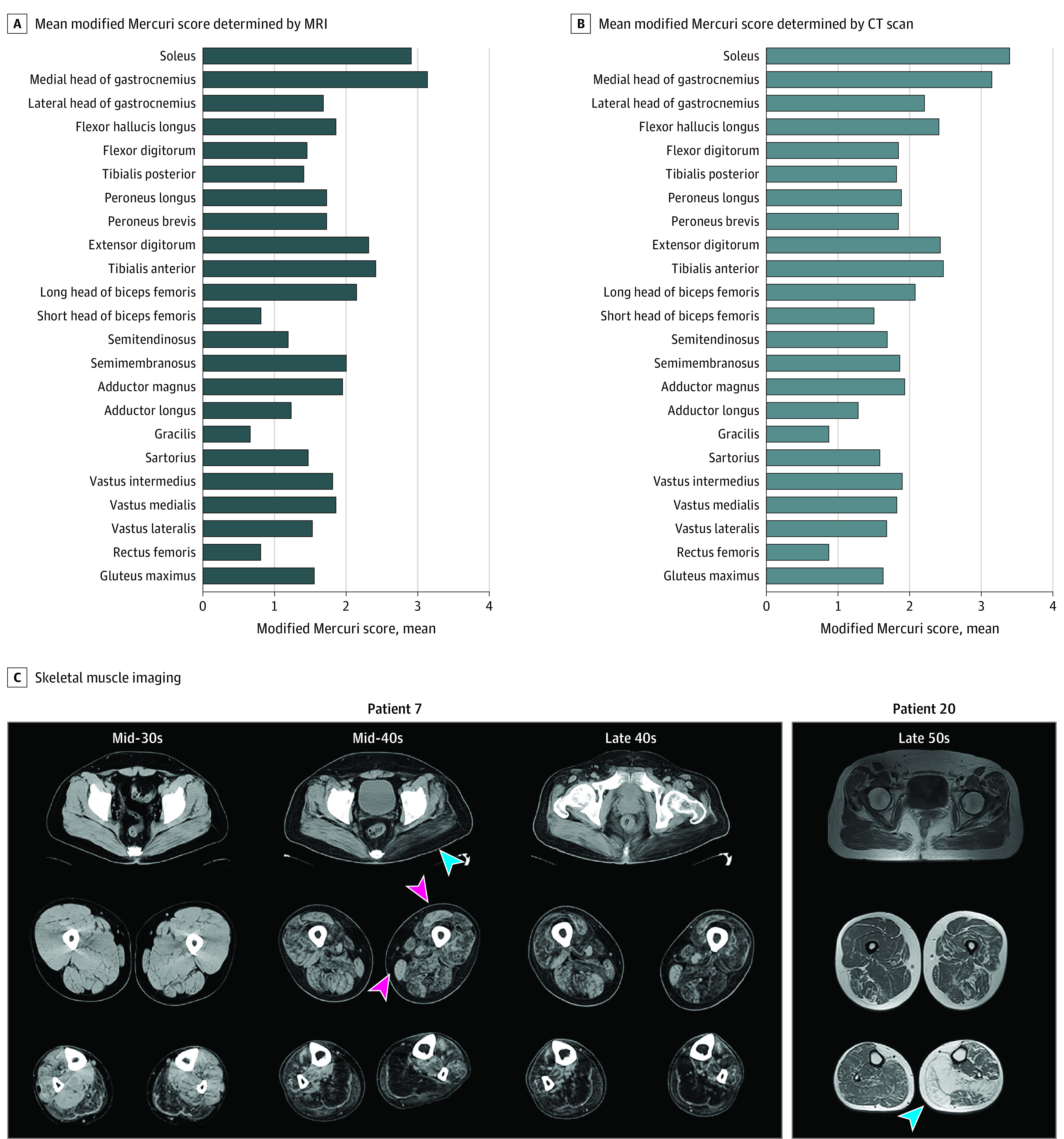
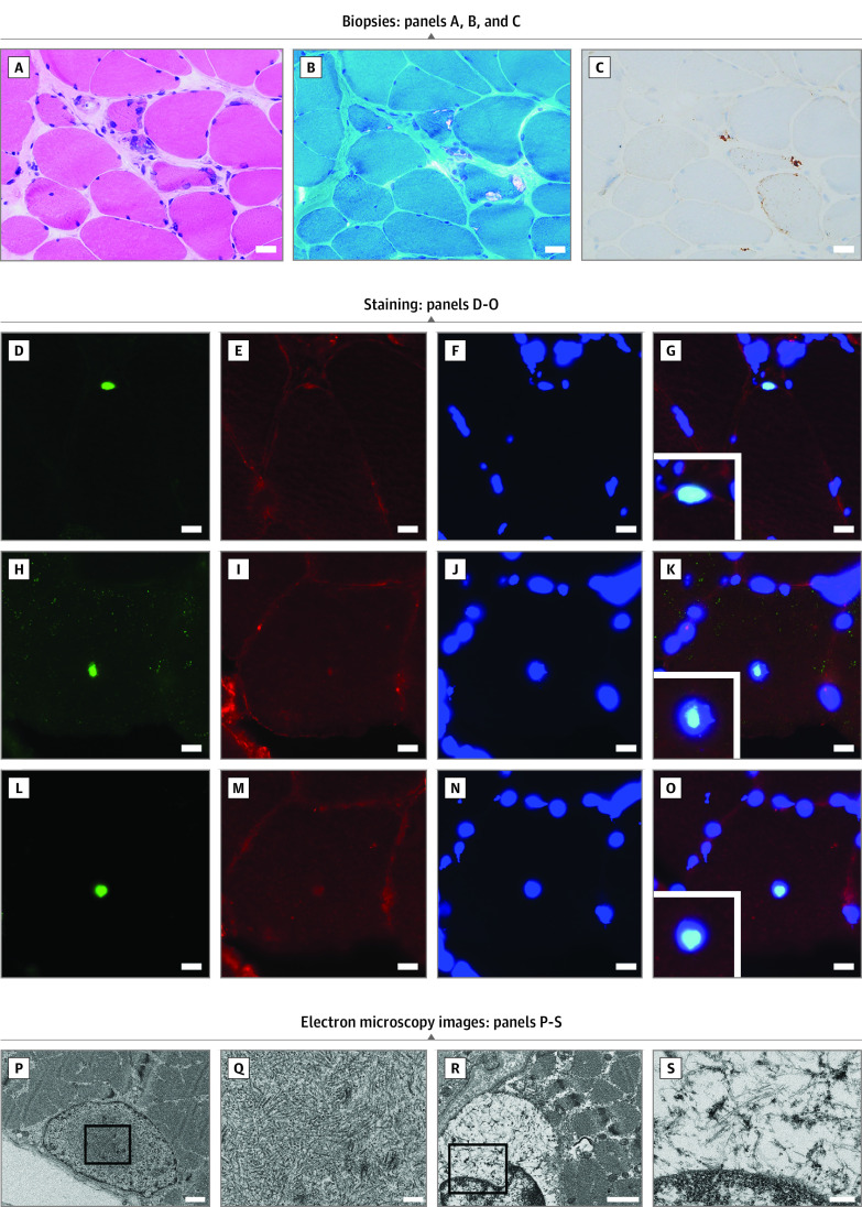
Results: Sixty-five Japanese patients with OPDM (40 men [62%]; mean [SD] age at onset, 41.0 [10.1] years) from 59 families with CGG repeat expansions in LRP12 were identified. This represents the most common OPDM subtype among all patients in Japan with genetically diagnosed OPDM. The expansions ranged from 85 to 289 repeats. A negative correlation was observed between the repeat size and the age at onset (r2 = 0.188, P = .001). The most common initial symptoms were ptosis and muscle weakness, present in 24 patients (37%). Limb muscle weakness was predominantly distal in 53 of 64 patients (83%), but 2 of 64 patients (3%) had predominantly proximal muscle weakness. Ptosis was observed in 62 of 64 patients (97%), and dysphagia or dysarthria was observed in 63 of 64 patients (98%). A total of 21 of 64 patients (33%) had asymmetric muscle weakness. Aspiration pneumonia was seen in 11 of 64 patients (17%), and 5 of 64 patients (8%) required mechanical ventilation. Seven of 64 patients (11%) developed cardiac abnormalities, and 5 of 64 patients (8%) developed neurologic abnormalities. Asymmetric muscle involvement was detected on computed tomography scans in 6 of 27 patients (22%) and on magnetic resonance imaging scans in 4 of 15 patients (27%), with the soleus and the medial head of the gastrocnemius being the worst affected. All 42 muscle biopsy samples showed rimmed vacuoles. Intranuclear tubulofilamentous inclusions were observed in only 1 of 5 patients.

Conclusions and relevance: This study suggests that OPDM_LRP12 is the most frequent OPDM subtype in Japan and is characterized by oculopharyngeal weakness, distal myopathy that especially affects the soleus and gastrocnemius muscles, and rimmed vacuoles in muscle biopsy.


Conflict of interest statement

Conflict of Interest Disclosures: Dr Ogasawara reported receiving grants from JSPS KAKENHI 20K16612 outside the submitted work. Dr Ishiura reported receiving grants from Japan Society for the Promotion of Science and The Kato Memorial Trust for Nambyo Research during the conduct of the study; and having a patent for diagnosis of OPDM pending. Dr S. Tsuji reported receiving grants from Nobelpharma Co Ltd; personal fees from Sanofi KK, Ono Pharmaceutical Co Ltd, and Sanwa Kagaku Kenkyusho Co Ltd outside the submitted work; and having a patent for diagnosis of OPDM pending. Dr Shimizu reported receiving grants from Tokyo Metropolitan Institute of Medical Science and JSPS KAKENHI by the Ministry of Education, Culture, Sports, Science and Technology of Japan outside the submitted work. Dr Kurashige reported receiving grants from Tsuchiya Foundation, Takeda Science Foundation, and Taiju Life Social Welfare Foundation outside the submitted work. Dr Noguchi reported receiving grants from Daiichi Sankyo Company and Astellas Pharma Inc outside the submitted work. Dr Nishino reported receiving honoraria from Sanofi and Japan Blood Products Organization; and a grant from Astellas Pharma Inc outside the submitted work. No other disclosures were reported.

Figures

Figure 1.
Figure 1.. Screening of the Patients
A total of 198 families with clinical or clinicopathologic diagnoses of oculopharyngodistal myopathy (OPDM) or oculopharyngeal muscular dystrophy (OPMD) were screened for CGG repeat expansion. Patients with GCN repeats in PABPN1 were excluded. Muscle biopsy samples were available from 112 families, and the samples from 65 families showed rimmed vacuoles. Among the samples with rimmed vacuoles, repeat expansion was identified in the samples from 54 families (42 in LRP12, 5 in GIPC1, and 7 in NOTCH2NLC). No sequence variant was identified in 47 families that did not show rimmed vacuoles in muscle biopsy samples. aPatients with OPDM_GIPC1 and OPDM_NOTCH2NLC have been previously reported and described.,
Figure 2.
Figure 2.. Genetic Analysis of Patients With OPDM_LRP12
A, Regression analysis between CGG repeat expansion size and the age at onset (r2 = 0.188; P = .001; n = 52). B, Pedigree of family 1. C, Southern blot analysis of family 1; patients 14 (II-3) and 27 (II-1) showed expansions of 144 and 140 CGG repeats, respectively. Their asymptomatic father (I-1) has mosaicism (arrows) and harbors 336 and 364 CGG repeats. Their mother (I-2) does not exhibit CGG expansion. D, Pedigree of family 2. E, Southern blot analysis of family 2; patients 57 (IV-2), 58 (III-2), and 59 (II-5) revealed mosaicism (219 and 289) and expansions of 85 and 127 repeats, respectively. The other 2 family members (III-4 and II-4) without symptoms harbor expansions of 63 and 49 repeats, respectively.
Figure 3.
Figure 3.. Muscle Imaging Results of Patients With OPDM_LRP12
A, Mean modified Mercuri scale score determined by T1-weighted magnetic resonance imaging (MRI) of 15 patients with OPDM_LRP12. B, Mean modified Mercuri scale score determined by computed tomography (CT) for 27 patients with OPDM_LRP12. C, Skeletal muscle imaging for patients with OPDM with LRP12 variations. Muscle imaging scans for patients 7 and 20 are shown. The distal muscles were affected earlier and more severely than the proximal muscles. Asymmetrical involvement was observed in the gluteus muscle and the lower legs (blue arrowheads). In the lower legs, the earliest and most severe involvement was observed in the soleus and gastrocnemius muscles. In the upper legs, the adductor magnus, semimembranosus, and short head of the biceps femoris were more affected, whereas the rectus femoris and gracilis muscles were usually preserved in patients with OPDM_LRP12 (pink arrowheads).
Figure 4.
Figure 4.. Histopathologic and Electron Microscopy Findings in Patients With OPDM_LRP12 Patients
A, B, and C, Biopsies from the left biceps brachii of patient 21, who has had the disease for 8 years, are shown. Scale bar: 20 μm. A, Moderate to marked fiber size variation and fibers with internal nuclei are seen on hematoxylin-eosin stain. B, Fibers with rimmed vacuole and moderate fibrous tissue infiltration are seen on modified Gomori trichrome stain. C, The dotlike deposition of p62 can be observed in muscle fibers. Staining with anti–SUMO-1 antibody (D), anti–caveolin-3 antibody (E, I, M), 4′,6-diamidino-2-phenylindole (F, J, N), anti-p62 (H), and anti–poly-ubiquitinated protein antibody (L). G, K, and O show merged immunohistochemistry. D to O, Scale bar: 10 μm. P and Q, Electron microscopy images of the biopsied left biceps brachii muscle from patient 10, in their late 60s. Scale bars are 1 μm for P and 200 nm for Q: Intranuclear tubulofilamentous inclusions (mean [SD] diameter, 17.3 [1.4] nm) are shown. R and S, Electron microscopy images of the biopsied left deltoid from patient 14, in their late 30s. Scale bars are 1 μm for R and 200 nm for S: Cytoplasmic tubulofilamentous inclusions (diameter, 10 nm) are shown.

Similar articles

Cited by

References

    1. Satoyoshi E, Kinoshita M. Oculopharyngodistal myopathy. Arch Neurol. 1977;34(2):89-92. doi:10.1001/archneur.1977.00500140043007 - DOI - PubMed
    1. Durmus H, Laval SH, Deymeer F, et al. . Oculopharyngodistal myopathy is a distinct entity: clinical and genetic features of 47 patients. Neurology. 2011;76(3):227-235. doi:10.1212/WNL.0b013e318207b043 - DOI - PubMed
    1. Minami N, Ikezoe K, Kuroda H, Nakabayashi H, Satoyoshi E, Nonaka I. Oculopharyngodistal myopathy is genetically heterogeneous and most cases are distinct from oculopharyngeal muscular dystrophy. Neuromuscul Disord. 2001;11(8):699-702. doi:10.1016/S0960-8966(01)00227-9 - DOI - PubMed
    1. Lu H, Luan X, Yuan Y, Dong M, Sun W, Yan C. The clinical and myopathological features of oculopharyngodistal myopathy in a Chinese family. Neuropathology. 2008;28(6):599-603. doi:10.1111/j.1440-1789.2008.00924.x - DOI - PubMed
    1. Zhao J, Liu J, Xiao J, et al. . Clinical and muscle imaging findings in 14 mainland Chinese patients with oculopharyngodistal myopathy. PLoS One. 2015;10(6):e0128629. doi:10.1371/journal.pone.0128629 - DOI - PMC - PubMed

Publication types

Substances

Supplementary concepts