. 2021 Dec;31(12):9038-9047.
doi: 10.1007/s00330-021-08013-6. Epub 2021 May 28.

Diffusion-weighted imaging diagnostic algorithm in patients with suspected pleural malignancy

Affiliations

Diffusion-weighted imaging diagnostic algorithm in patients with suspected pleural malignancy

Wenrui Jiang et al. Eur Radiol. 2021 Dec.

Abstract

Objectives: The purpose of this study was to analyze the diagnostic performance and clinical application of diffusion-weighted imaging (DWI) in patients with suspected pleural malignancy (PM).

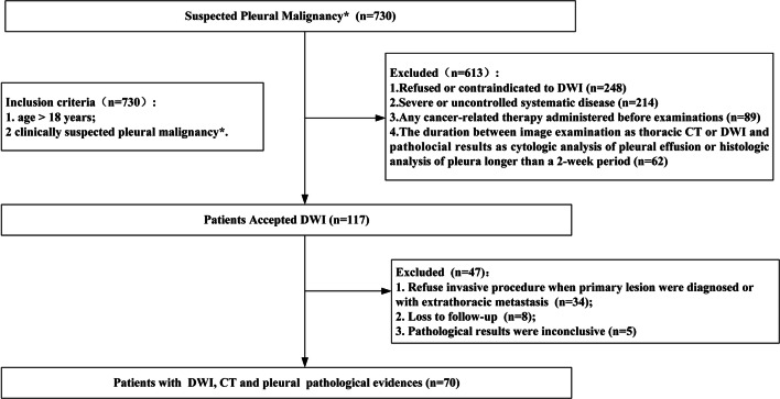
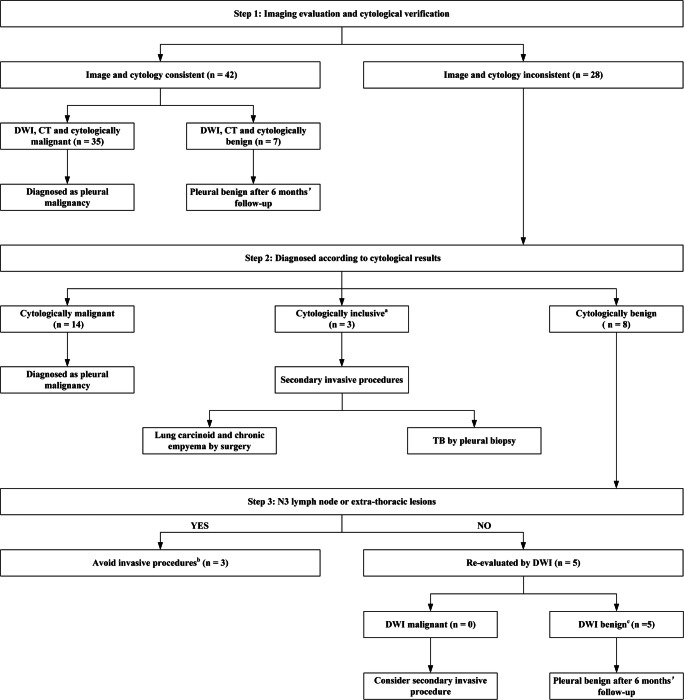
Methods: A retrospective review of patients with suspected PM was performed from March 2014 to August 2018 (NCT02320617). All patients underwent chest DWI and computed tomography (CT) with cytological or histopathological findings as reference standards. The diagnostic performance of DWI and CT was analyzed and compared. A DWI diagnostic algorithm with three sequential steps was established.

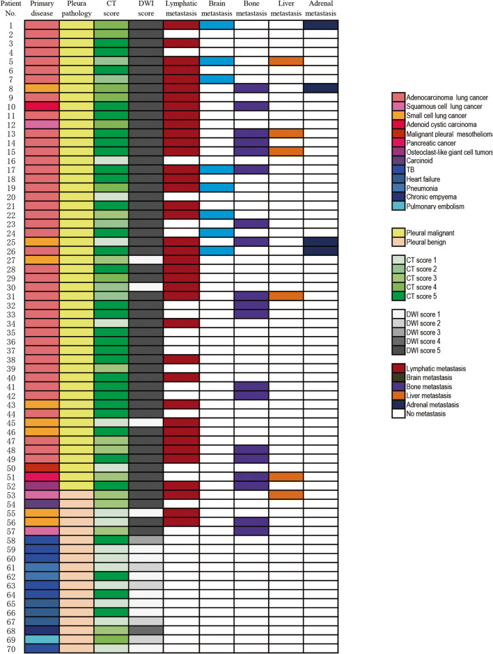
Results: Seventy patients (61.6 ± 13.6 years; 47 males and 23 females) were included. The sensitivity of DWI (94.2%, 49/52) for the diagnosis of PM was significantly higher compared with CT (67.3%, 35/52), with similar specificity (72.2% vs. 72.2%, respectively). The apparent diffusion coefficient of malignant lesions (1.15 ± 0.32 × 10⁻³ mm2/s) was lower compared with benign lesions (1.46 ± 0.68 × 10⁻³ mm2/s), but the cutoff value was difficult to define for overlap between groups. Approximately 62.5% (5/8) of invasive procedures were avoided when using the DWI diagnostic algorithm in patients with suspected PM without N3 lymph node or extra-thoracic metastasis.

Conclusion: Including DWI into the diagnostic algorithm of suspected PM can effectively identify malignancy and avoid unnecessary invasive procedures, which may have some potential in clinical application.

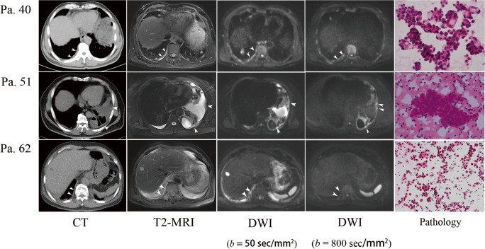
Key points: • Diffusion-weighted imaging can identify pleural malignancy much more efficiently than CT. • A diffusion-weighted imaging diagnostic algorithm helped to avoid unnecessary invasive procedures in patients without N3 lymph node or extra-thoracic lesions. • A hyperintense signal on DWI at a high b value (800 s/mm2) but not at a low b value (50 s/mm2) was a reliable signature of PM.

Keywords: Diffusion magnetic resonance imaging; Pleural neoplasms; Tomography, X-ray computed.


Conflict of interest statement

The authors of this manuscript declare no relationships with any companies whose products or services may be related to the subject matter of the article.

Figures

Fig. 1
Fig. 1
Flowchart of patient inclusion, exclusion, and recruitment. DWI: diffusion-weighted imaging. *Suspected pleural malignancy: (1) new effusion in patients with primary malignant disease; (2) any unexplained pleural lesion or effusion after primary care
Fig. 2
Fig. 2
Patient number and diagnosis of primary disease, pleural pathology, visual score on CT and DWI, and metastasis in lymph nodes, brain, bone, liver, and adrenal glands. TB: tuberculosis
Fig. 3
Fig. 3
Representative pleural malignant features on CT (ae) and DWI (fj) are marked by the white arrow: a circumferential pleural thickening; b nodular pleural thickening; c visceral pleural thickening > 1 cm; d mediastinal pleural involvement; e chest wall invasion and rib destruction at multiple sites; f circumferential pleural thickening; g nodular pleural thickening; h thready pleural thickening; i mediastinal pleural involvement; j chest wall invasion and rib destruction at multiple sites
Fig. 4
Fig. 4
DWI diagnostic algorithm of suspected PM. (a) Cytological inclusion was defined as atypia cells in pleural effusion. (b) Invasive procedures were avoided in these patients because management would change marginally with the diagnosis of PM. (c) Although PM features on CT were observed in these patients, a benign diagnosis was supported by both DWI and cytological reports and confirmed after 6 months of follow-up. TB: tuberculosis; DWI: diffusion-weighted imaging; PM: pleural malignancy; CT: computed tomography
Fig. 5
Fig. 5
Representative cases. Patient 40 was a 60-year-old male diagnosed with primary adenocarcinoma. Circumferential pleural thickening was observed on CT and reported as PM. Similar features were observed on T2-weighted MRI. Multiple hyperintense areas were observed on DWI at a low b value (50 s/mm2) and a high b value (800 s/mm2), and definite malignancy was concluded. The cytological results confirmed adenocarcinoma. Patient 51 was a 63-year-old female diagnosed with advanced adenocarcinoma with bone and liver metastasis. CT was equivocal in the pleural assessment. Thready pleural thickening was observed on T2-weighted MRI. Multiple hyperintense areas were present on DWI, especially at a high b value (800 s/mm2), and definite malignancy was concluded. Cytological results confirmed adenocarcinoma. Patient 62 was a 67-year-old male with recurrent pleural effusion. Circumferential pleural thickening was observed on CT, and definite malignancy was concluded. However, hyperintense areas present on DWI at a low b value (50 s/mm2) were not present at a high b value (800 s/mm2); thus, the patient was classified as benign. Benign results were reported by cytology, which was repeated three times, and effusion was completely resolved after antibiotic therapy and 6 months of follow-up.

References

    1. Goldstraw P, Chansky K, Crowley J, et al. The IASLC Lung Cancer Staging Project: proposals for revision of the TNM stage groupings in the forthcoming (Eighth) edition of the TNM classification for lung cancer. J Thorac Oncol. 2016;11(1):39–51. doi: 10.1016/j.jtho.2015.09.009. - DOI - PubMed
    1. Eberhardt WE, Mitchell A, Crowley J, et al. The IASLC Lung Cancer Staging Project: proposals for the revision of the M Descriptors in the forthcoming eighth edition of the TNM classification of lung cancer. J Thorac Oncol. 2015;10(11):1515–1522. doi: 10.1097/JTO.0000000000000673. - DOI - PubMed
    1. Ou SH, Zell JA. Validation study of the proposed IASLC staging revisions of the T4 and M non-small cell lung cancer descriptors using data from 23,583 patients in the California Cancer Registry. J Thorac Oncol. 2008;3(3):216–227. doi: 10.1097/JTO.0b013e318164545d. - DOI - PubMed
    1. Du Rand I, Maskell N. Introduction and methods: British Thoracic Society Pleural Disease Guideline 2010. Thorax. 2010;65 Suppl 2:ii1–ii3. doi: 10.1136/thx.2010.137042. - DOI - PubMed
    1. Arenas-Jimenez J, Alonso-Charterina S, Sanchez-Paya J, Fernandez-Latorre F, Gil-Sanchez S, Lloret-Llorens M. Evaluation of CT findings for diagnosis of pleural effusions. Eur Radiol. 2000;10(4):681–690. doi: 10.1007/s003300050984. - DOI - PubMed

LinkOut - more resources