Review
. 2021 Sep;9(5):1306-1321.
doi: 10.1111/andr.13056. Epub 2021 Jun 11.

The role of scrotal ultrasonography from infancy to puberty

Affiliations
Review

The role of scrotal ultrasonography from infancy to puberty

Matteo Spaziani et al. Andrology. 2021 Sep.

Abstract

Background: Scrotal ultrasonography is an essential diagnostic tool in daily clinical practice. The availability of new-generation ultrasound machines characterized by clearly improved image quality, low health cost, and higher patient safety, represents only some characteristics of ultrasound investigation. The usefulness of scrotal ultrasonography is particularly evident in the period of life from infancy to puberty, during which males undergo important morphofunctional changes, and several pathological conditions may occur.

Objectives: This pictorial review primarily aimed to investigate the aspects of ultrasonography related to the normal physiological development of the gonads from mini-puberty to pubertal onset. This study also aimed to provide an update on the use of ultrasonography in main andrological pathologies that may occur during this period. The conditions that are discussed in depth are: cryptorchidism, inguinoscrotal hernias, and hydrocele in the neonatal phase; acute scrotum, epididymo-orchitis, and testicular cancers in childhood; and hypogonadism, varicoceles, testicular microlithiasis, and oncohematological pathology in puberty.

Discussion: We provided an ultrasound slant for all the above-mentioned pathologies while purposely avoiding excessive deepening of the pathogenetic, clinical, and therapeutic aspects. Studying the ultrasound aspects of the gonads also facilitates differential diagnosis between various conditions and represents a good aid in evaluating therapeutic success (e.g., in hypogonadism or postsurgical evaluation of varicoceles and cryptorchidism).

Conclusion: Scrotal ultrasonography is now globally recognized as the necessary completion of clinical-laboratory overview in gonads evaluation. This diagnostic procedure is even more indispensable in the infancy-childhood-puberty period for the evaluation of normal gonadal development as well as diagnosis of other possible diseases.

Keywords: andrological pathologies; childhood; infancy; mini-puberty; puberty; scrotal ultrasonography.


Conflict of interest statement

The authors declare no conflict of interest.

Figures

FIGURE 1
FIGURE 1
Ultrasound image of the normal male testicle during mini‐puberty using high‐frequency (12 MHz) linear probe. During this phase of life, the testes appear symmetrical, equal in size (approximately 0.5–1.5 mL), and homogeneous, but also more hypoechoic than adult testes. Note that the mediastinum appears evident and tendentially hyperechogenic
FIGURE 2
FIGURE 2
Ultrasound images of the normal male testicle during childhood using high‐frequency (12 MHz) linear probe. (A)The testes appear symmetrical, equal in size (approximately 1.5–2 mL) and homogeneous. (B) An initial increase of the color signal is possible
FIGURE 3
FIGURE 3
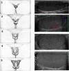
Clinical progression of pubertal stages from G1 to G5 with related ultrasound modifications. It is possible to note the increase in volume and echogenicity as pubertal stages and seminiferous tubule maturation advance
FIGURE 4
FIGURE 4
Ultrasound images of an inguinal cryptorchid testes using high‐frequency (12 MHz) linear probe. The testicle appears hypoechoic
FIGURE 5
FIGURE 5
Ultrasound images of a neonatal hydrocele using high‐frequency (12 MHz) linear probe. The testes are surrounded by anechoic fluid collection, with the posterior margin firmly adherent to epididymis and scrotal walls
FIGURE 6
FIGURE 6
Ultrasound images of (A, B) testicular and (C, D) epididymal appendages using high‐frequency (12 MHz) linear probe. They appear oval in shape and isohypoechoic; visualization may be aided by the presence of a reactive hydrocele
FIGURE 7
FIGURE 7
High‐frequency (12 MHz) Doppler ultrasound images of testicular epidermoid cysts from two 17‐year‐old pubertal boys. (A) The typical onion ring appearance presentation of a solid mass surrounded by concentric rings of hypo‐ and hyperechogenicity. (B) Another epidermoid cyst characterized by a particularly calcific margin that generates an evident posterior shadow cone. Scattered microlithiasis is also visible
FIGURE 8
FIGURE 8
Ultrasound images of testicular recurrence of leukemia using high‐frequency (12 MHz) linear probe. (A) Several hypoechogenic foci of leukemic infiltration (recurrence of acute lymphatic leukemia). (B) The color Doppler US results, which shows an increased blood flow in each lesion
FIGURE 9
FIGURE 9
Ultrasound images of testis appearance in Klinefelter syndrome using high‐frequency (12 MHz) linear probe. (A) Some foci of Leydig cell hyperplasia as hypoechoic round lesion with regular but blurred margins; (B) the different ultrasound histological damage aspects of the gonads during the succession of pubertal stages. The parenchymal irregular hypoechogenicity and hypervascularization are particularly evaluable. The last image reports a focus of Leydig cell hyperplasia
FIGURE 10
FIGURE 10
High‐frequency (12 MHz) linear probe ultrasound images of testicular varicocele. (A, B) Normal grayscale images, (C, D) color Doppler analysis
FIGURE 11
FIGURE 11
High‐frequency (12 MHz) ultrasound images of testicular microlithiasis. (A, B) Infant patients, (C) a pubertal boy with a classic “starry sky” appearance

Similar articles

Cited by

References

    1. Becker M, Hesse V. Minipuberty: why does it happen? Horm Res Paediatr. 2020;93(2):76–84. - PubMed
    1. Kuiri‐Hänninen T, Sankilampi U, Dunkel L. Activation of the hypothalamic‐pituitary‐gonadal axis in infancy: minipuberty. Horm Res Paediatr. 2014;82(2):73–80. - PubMed
    1. Marshall WA, Tanner JM. Variations in the pattern of pubertal changes in boys. Arch Dis Child. 1970;45(239):13–23. - PMC - PubMed
    1. Spaziani M, Tarantino C, Tahani N, et al. Hypothalamo‐Pituitary axis and puberty. Mol Cell Endocrinol. 2021;520:111094. - PubMed
    1. Delaney LR, Karmazyn B. Ultrasound of the pediatric scrotum. Semin Ultrasound CT MR. 2013;34(3):248–256. 10.1053/j.sult.2012.11.010 - DOI - PubMed

Publication types