. 2021 May 1;22(5):1623-1632.
doi: 10.31557/APJCP.2021.22.5.1623.

Rosmarinic Acid Induces Proliferation Suppression of Hepatoma Cells Associated with NF-κB Signaling Pathway

Affiliations

Rosmarinic Acid Induces Proliferation Suppression of Hepatoma Cells Associated with NF-κB Signaling Pathway

Yanjun An et al. Asian Pac J Cancer Prev. .

Abstract

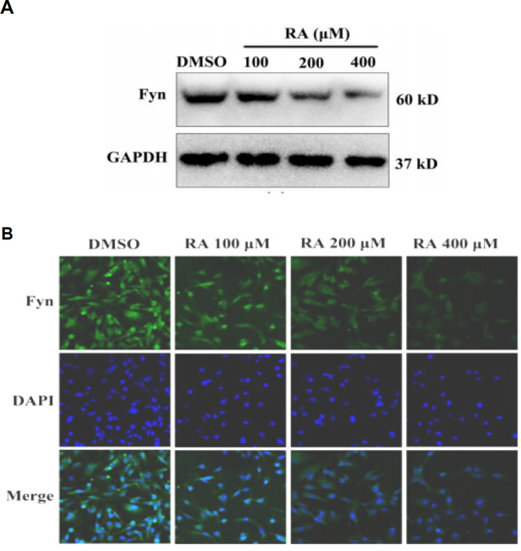
Background: Rosmarinic acid (RA) is a natural phenolic compound that acts as a Fyn inhibitor by 53 homology modeling of the human Fyn structure. Therefore, the apoptosis mechanism related to NF-κB signaling pathway induced by RA in HepG2 was investigated.

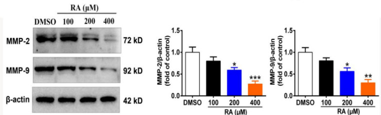
Methods: The cell growth, apoptosis, and proliferation of HepG2 regulated by various concentrations of RA were studied. The proteins expression of MMP-2, MMP-9, PI3K, AKT, NF-κB, and apoptosis-related proteins Bax, Bcl-2, cleaved caspase-3 were detected.

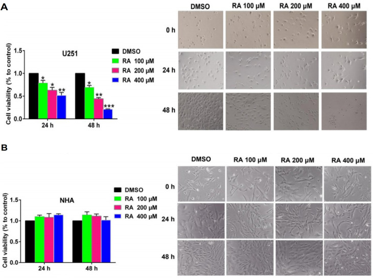
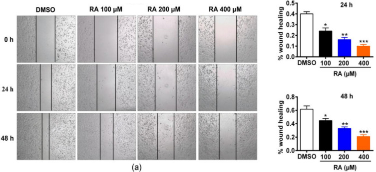
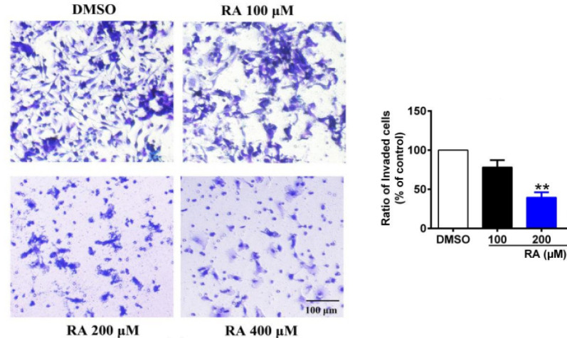
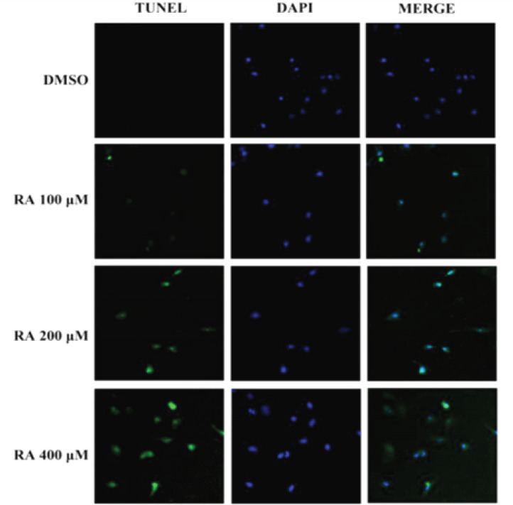
Results: RA significantly reduced proliferation rates, inhibited migration and invasion, and decreased the expressions of invasion-related factors, such as matrix metalloproteinase (MMP)-2 and MMP-9. TUNEL staining revealed that RA resulted in a dose-dependent increase of HepG2 cell apoptosis. In line with this finding, the expression of apoptosis suppressor protein Bcl-2 was downregulated and that of the pro-apoptotic proteins Bax and cleaved caspase-3 was increased. In addition, we found that the phosphatidylinositol 3-kinase (PI3K)/Akt/nuclear factor kappa B (NF-κB) signaling pathway was involved in RA-mediated inhibition of HepG2 cell metastasis.

Conclusion: Our study identified that RA as a drug candidate for the treatment of HCC.

Keywords: HepG2 cells; Proliferation; Rosmarinic acid (RA); hepatocellular carcinoma (HCC).


Conflict of interest statement

All authors declares that we have no conflict of interest.

Figures

Figure 1
Figure 1
RA Inhibits Expression of Fyn. We firstly examined the effect of RA on the expression of Fyn in HepG2 cells. RA treatment reduced Fyn expression in HepG2 cells in a dose-dependent manner (A). Immunofluorescence staining confirmed that RA inhibits Fyn expression in HepG2 cells (B)
Figure 2
Figure 2
RA Inhibits Hepatoma Cell Proliferation. Next, we examined the effect of RA on hepatoma cell proliferation using the CCK-8 assay. RA treatment inhibited cell proliferation in a time- and dose-dependent manner in HepG2 cells. RA-induced cytotoxicity was also observed under a microscope. The images show that as the concentration of RA increased, the density of hepatoma cells decreased. Moreover, the cells shrank and died more. These observations were in line with the results of the CCK-8 assay (A). As a control, we also examined the effect of RA on NHA. RA treatment for 24 hrs or 48 hrs did not significantly affect NHA cell viability and morphology (B). These results indicated that RA specifically inhibited the proliferation of hepatoma cells, with no significant effect on the proliferation of NHA
Figure 3
Figure 3
RA Inhibits Hepatoma Cell Migration. Metastasis of rapidly migrating tumor cells is the main cause of death for most patients with cancer [19]. Therefore, inhibiting migration could be an important strategy to prevent tumor metastasis. We examined the effect of RA on hepatoma cell migration. HepG2 cells showed reduced migration after treatment with RA for 24 hrs or 48 hrs at different concentrations. Specifically, in the control group of the HepG2 cell line, wound healing reached 40% after 24 hrs, while it only reached 24%, 18%, and 10% after treatment with 100, 200, and 400 µM RA, respectively. At 48 hrs, wound healing of control HepG2 cells reached 60%, but 42%, 30%, and 20% after treatment with 100, 200, and 400 µM RA, respectively
Figure 4
Figure 4
RA Inhibits Hepatoma Cell Invasion. The high invasion capability of hepatoma cells is the main reason for the high refractory and recurrence rates of HCC. Therefore, we tested the effect of RA on hepatoma cell invasion using a Matrigel® invasion assay. RA significantly inhibited invasion through the Matrigel®. Compared with the control group, the percentage of invading HepG2 cells was decreased by 21.7%, 60.3%, and 76.4% after treatment with 100, 200, and 400 µM RA for 24 hrs, respectively
Figure 5
Figure 5
RA Inhibits Hepatoma Cell Invasion. Moreover, we detected the expression of MMP-2 and MMP-9 by western blot. RA dose-dependently inhibited the expression of MMP-2 and MMP-9 in HepG2 cells. Compared with the control group, the expression of MMP-2 in HepG2 cells was decreased by 20.0%, 40.7%, and 62.7%, and that of MMP-9 was decreased by 19.9%, 43.7%, and 70%, after treatment with 100, 200, and 400 µM for 24 h, respectively. These results suggested that RA inhibited hepatoma cell invasion and migration by reducing the expression of MMPs, thereby providing pathways for the invasion and metastasis of hepatoma cells
Figure 6
Figure 6
RA Increases Hepatoma Cell Apoptosis. We investigated whether RA could induce apoptosis. TUNEL assay revealed that RA induced apoptosis significantly in a dose-dependent manner in HepG2 cells
Figure 7
Figure 7
RA Increases Hepatoma Cell Apoptosis. We found that RA treatment increased the expression of cleaved caspase-3 and Bax and reduced the expression of Bcl-2 in HepG2 cells. In addition, RA treatment increased the relative ratio of Bax/Bcl-2 as well as cleaved caspase3/caspase3, indicating RA-induced apoptosis in hepatoma cells. Compared with the control group, the relative ratio of Bax/Bcl-2 in HepG2 cells increased 7.6%, 63.0%, and 156.3%, after treatment with 100, 200, and 400 µM RA for 24 hrs, respectively. In addition, the relative ratio of cleaved caspase3/caspase3 in HepG2 cells increased 27.3%, 79.6%, and 124.3%, after treatment with 100, 200, and 400 µM RA for 24 hrs
Figure 8
Figure 8
RA Inhibits the PI3K/Akt/NF-κB Signaling Pathway in Hepatoma Cells. We examined the effect of RA on the PI3K/Akt/NF-κB signaling pathway. We found that treatment with 100, 200, and 400 µM RA for 24 hrs dramatically decreased the protein expression of PI3K, p-Akt, and NF-κB in HepG2 cells compared to control cells. Specifically, the expression of PI3K was decreased by 11.7%, 24%, and 50.3% in HepG2 cells, respectively. In HepG2 cells, the expression of p-Akt was decreased by 18.0%, 51.7%, and 66.7% and that of NF-κB p65 by 30.7%, 31.4%, and 56%, respectively. Collectively, these results suggested that RA might inhibit proliferation, migration, and invasion and induce apoptosis of hepatoma cells via the PI3K/Akt/NF-κB signaling pathway

References

    1. Affo S, Yu LX, Schwabe RF. The role of cancer-associated fibroblasts and fibrosis in liver cancer. Annu Rev Pathol. 2017;12:153–86. - PMC - PubMed
    1. Alcaraz M, Alcaraz-Saura M, Achel DG, et al. Radiosensitizing effect of rosmarinic acid in metastatic melanoma B16F10 cells. Anticancer Res. 2014;34:1913–21. - PubMed
    1. Bosetti C, Turati F, La Vecchia C. Hepatocellular carcinoma epidemiology. Best Pract Res Clin Gastroenterol. 2014;28:753–70. - PubMed
    1. Camargo ES, Viveiros MM, Correa NI, Robles L, Rezende MB. Primary hepatic carcinoid tumor: case report and literature review. Einstein (Sao Paulo) 2014;12:494–508. - PMC - PubMed
    1. Cheng JC, Chou CH, Kuo ML, Hsieh CY. Radiation-enhanced hepatocellular carcinoma cell invasion with MMP-9 expression through PI3K/Akt/NF-kappaB signal transduction pathway. Oncogene. 2006;25:7009–18. - PubMed

MeSH terms