

Review
doi: 10.1259/bjr.20201290.
Epub 2021 May 28.
Spectral detector CT applications in advanced liver imaging
Affiliations
- PMID: 34048285
- PMCID: PMC8248211
- DOI: 10.1259/bjr.20201290
Item in Clipboard
Review
Spectral detector CT applications in advanced liver imaging
Br J Radiol.
.
Abstract
Objective: Spectral detector CT (SDCT) has many applications in advanced liver imaging. If appropriately utilized, this technology has the potential to improve image quality, provide new diagnostic information, and allow for decreased radiation dose. The purpose of this review is to familiarize radiologists with the uses of SDCT in liver imaging.
Conclusion: SDCT has a variety of post-processing techniques, which can be used in advanced liver imaging and can significantly add value in clinical practice.
Figures
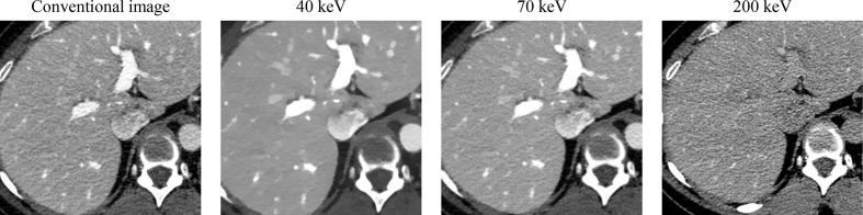
46-year-old male undergoing preoperative CT for potential liver donation. At 40 keV, there is increased iodine conspicuity in comparison with the conventional image. 70 keV image is equivalent to conventional image at 120 kVp in terms of iodine contrast. At 200 keV, iodine conspicuity decreases.
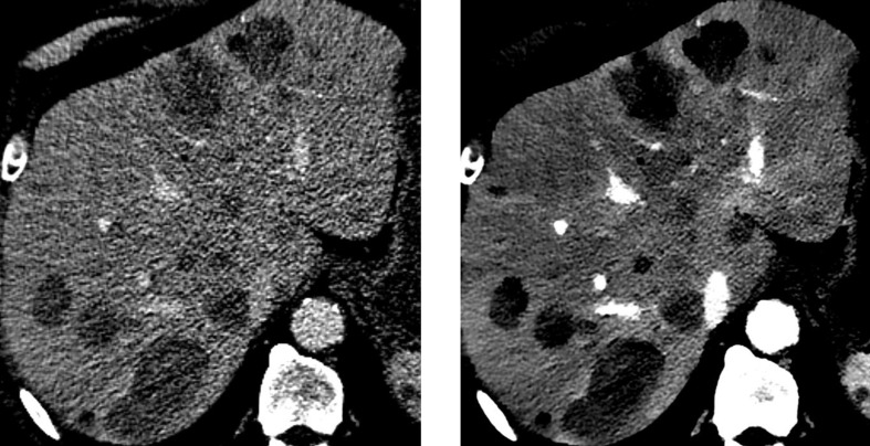
81-year-old male with pancreatic neuroendocrine tumor. There is increased conspicuity of hypovascular liver metastases on 40 keV image (right) compared with conventional image (left).

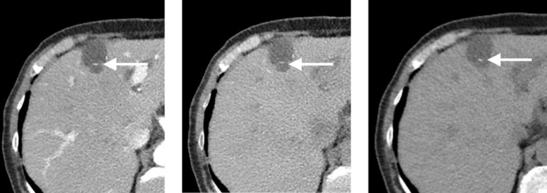
66-year-old male with cirrhosis. There is increased conspicuity of hypervascular liver lesion (arrows) on 50 keV image (right) compared with conventional image (left). This was subsequently diagnosed as hepatocellular carcinoma.
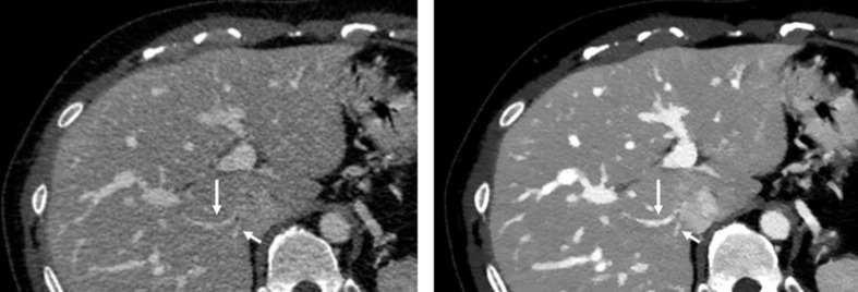
46-year-old male undergoing preoperative CT for potential liver donation. Accessory hepatic veins (arrows) are easier to visualize on 50 keV image (right) compared with conventional image (left).
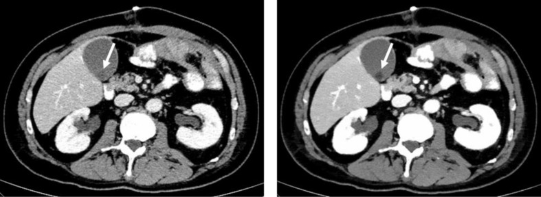
53-year-old male status post bowel resection; CT scan was performed to rule out intra-abdominal abscess. Incidentally noted is a noncalcified gallstone (arrows) that is more conspicuous on 50 keV image (right) compared with conventional image (left).
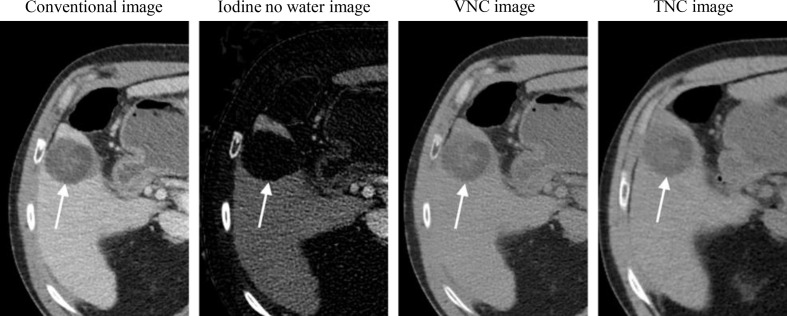
64-year-old male with cirrhosis, 5 months status post microwave ablation of segment five hepatocellular carcinoma. Zone of ablation (arrows) contains areas of hyperattenuation on conventional image. However, no iodine is visualized within the zone on iodine no water image. Hyperattenuation remains on virtual non-contrast (VNC) image which appears similar totrue non-contrast (TNC) image.
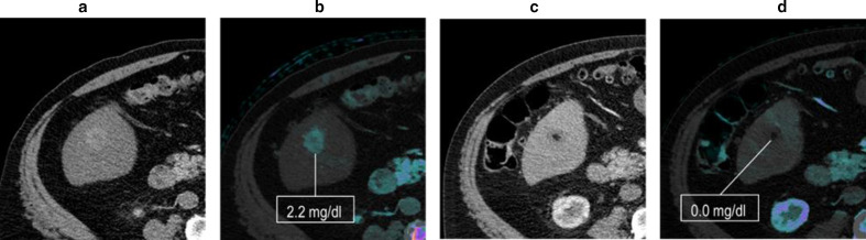
70-year-old male with cirrhosis and hepatocellular carcinoma, pre-treatment (A-B) and 3 months status post-TACE (C-D). (A) Conventional image showing arterially enhancing lesion in segments 5/6. (B) The lesion is demonstrated on an iodine overlay, with a measured iodine concentration of 2.2 mg dl−1. (C) Following ablation, on conventional image, there is some peripheral high density in the ablation zone; it is unclear if this represents hemorrhagic debris or residual disease. (D) The absence of iodine is confirmed on iodine overlay with a measured iodine concentration of 0 mg dl−1. This allows confident exclusion of residual or recurrent disease.
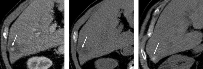
73-year-old male with hepatocellular carcinoma status post transarterial chemoembolization. Hyperdensity is seen within the lesion on arterial phase image (left), which suppresses on the VNC image (middle). This could have been misinterpreted as enhancement. However, TNC image (right) from a recent examination demonstrated the same intralesional hyperdensity, suggesting lipiodol deposition rather than enhancement.
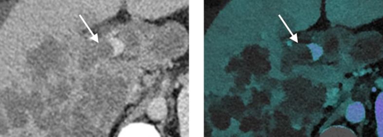
48-year-old male with infiltrative hepatocellular carcinoma. Conventional image (left) demonstrates infiltrative hepatocellular carcinoma and expansile thrombus within the right portal vein. Faint iodine is seen within the thrombus on iodine overlay image (right), favoring tumor thrombus.
87-year-old female with an incidentally visualized hepatic cyst containing a calcified septation (arrows). Conventional contrast enhanced image (left) demonstrates a cyst with a hyperdense septation. VNC image (middle) demonstrates persistent high attenuation focus within the septation, confirming that this represents calcification instead of enhancement. Calcification is confirmed on the TNC image (right).
Similar articles
-
Thoracic-abdominal imaging with a novel dual-layer spectral detector CT: intra-individual comparison of image quality and radiation dose with 128-row single-energy acquisition.Acta Radiol. 2018 Dec;59(12):1458-1465. doi: 10.1177/0284185118762611. Epub 2018 Mar 23. Acta Radiol. 2018. PMID: 29569933
-
Value of virtual monochromatic spectral image of dual-layer spectral detector CT with noise reduction algorithm for image quality improvement in obese simulated body phantom.BMC Med Imaging. 2019 Aug 28;19(1):76. doi: 10.1186/s12880-019-0367-8. BMC Med Imaging. 2019. PMID: 31462212 Free PMC article.
-
Quality of routine diagnostic abdominal images generated from a novel detector-based spectral CT scanner: a technical report on a phantom and clinical study.Abdom Radiol (NY). 2017 Nov;42(11):2752-2759. doi: 10.1007/s00261-017-1170-z. Abdom Radiol (NY). 2017. PMID: 28493070
-
Abdominal Applications of a Novel Detector-Based Spectral CT.Curr Probl Diagn Radiol. 2018 Mar-Apr;47(2):110-118. doi: 10.1067/j.cpradiol.2017.05.001. Epub 2017 May 17. Curr Probl Diagn Radiol. 2018. PMID: 28673603 Review.
-
The optimal contrast media policy in CT of the liver. Part I: Technical notes.Acta Radiol. 2011 Jun 1;52(5):467-72. doi: 10.1258/ar.2011.100499. Epub 2011 Mar 17. Acta Radiol. 2011. PMID: 21498281 Review.
Cited by
-
Intra-patient variability of iodine quantification across different dual-energy CT platforms: assessment of normalization techniques.Eur Radiol. 2024 Aug;34(8):5131-5141. doi: 10.1007/s00330-023-10560-z. Epub 2024 Jan 8. Eur Radiol. 2024. PMID: 38189979
-
Myocardial extracellular volume fraction with spectral detector computed tomography for risk stratification in non-ischemic heart failure.Radiol Med. 2025 Jul;130(7):1092-1104. doi: 10.1007/s11547-025-02002-1. Epub 2025 Apr 30. Radiol Med. 2025. PMID: 40304953 Free PMC article.
-
Application of Dual-Layer Spectral-Detector Computed Tomography Angiography in Identifying Symptomatic Carotid Atherosclerosis: A Prospective Observational Study.J Am Heart Assoc. 2024 Mar 19;13(6):e032665. doi: 10.1161/JAHA.123.032665. Epub 2024 Mar 18. J Am Heart Assoc. 2024. PMID: 38497470 Free PMC article.
-
Dual-layer spectral CT fusion imaging for lung biopsies: more accurate targets, diagnostic samplings, and biomarker information?Eur Radiol Exp. 2022 Aug 15;6(1):34. doi: 10.1186/s41747-022-00290-0. Eur Radiol Exp. 2022. PMID: 35965267 Free PMC article.
-
Noninvasive diagnosis of liver cirrhosis: qualitative and quantitative imaging biomarkers.Abdom Radiol (NY). 2024 Jun;49(6):2098-2115. doi: 10.1007/s00261-024-04225-8. Epub 2024 Feb 19. Abdom Radiol (NY). 2024. PMID: 38372765 Review.
References
-
- Hojjati M, Van Hedent S, Rassouli N, Tatsuoka C, Jordan D, Dhanantwari A, et al. . Quality of routine diagnostic abdominal images generated from a novel detector-based spectral CT scanner: a technical report on a phantom and clinical study. Abdom Radiol 2017; 42: 2752–9. doi: 10.1007/s00261-017-1170-z - DOI - PubMed
Publication types
MeSH terms
Substances
LinkOut - more resources
Full Text Sources
Other Literature Sources
Medical
