. 2021 May 28;17(5):e1009630.
doi: 10.1371/journal.ppat.1009630. eCollection 2021 May.

An incoherent feedforward loop formed by SirA/BarA, HilE and HilD is involved in controlling the growth cost of virulence factor expression by Salmonella Typhimurium

Affiliations

An incoherent feedforward loop formed by SirA/BarA, HilE and HilD is involved in controlling the growth cost of virulence factor expression by Salmonella Typhimurium

Deyanira Pérez-Morales et al. PLoS Pathog. .

Abstract

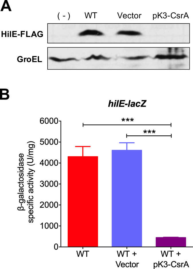
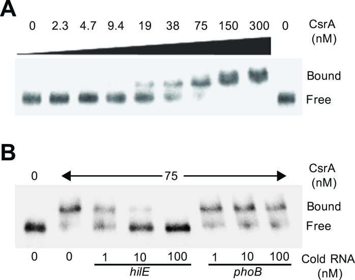
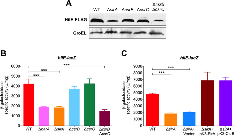
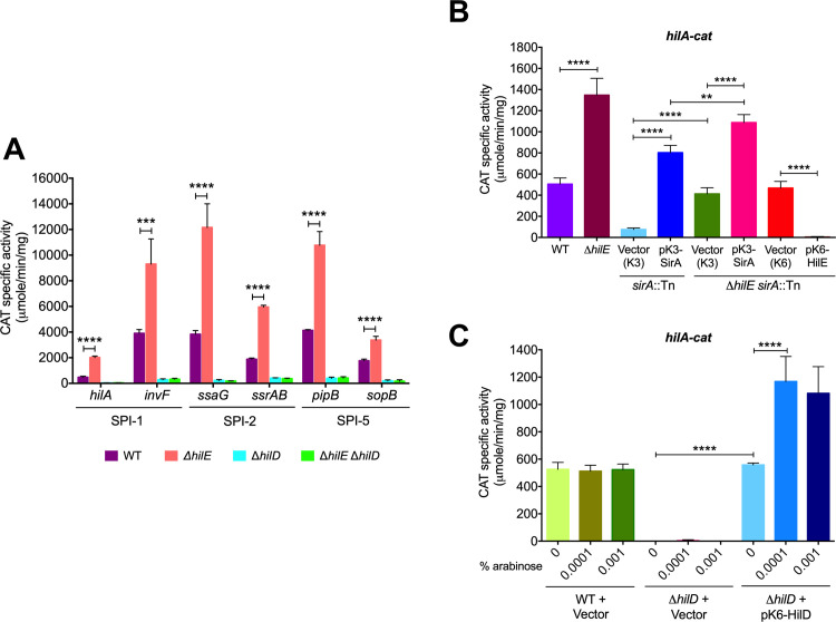
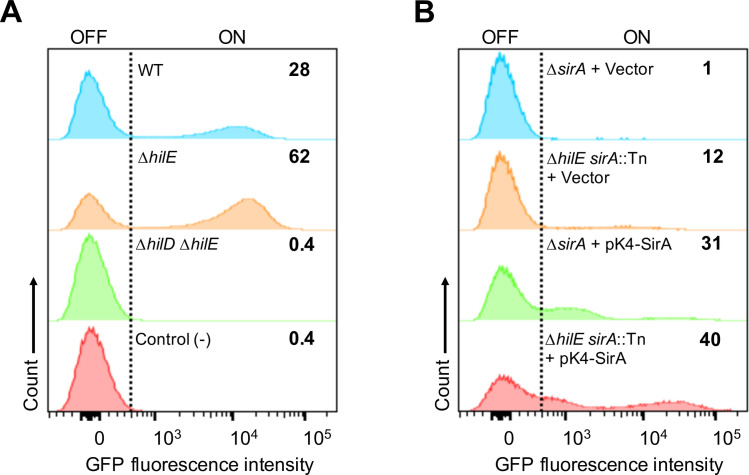
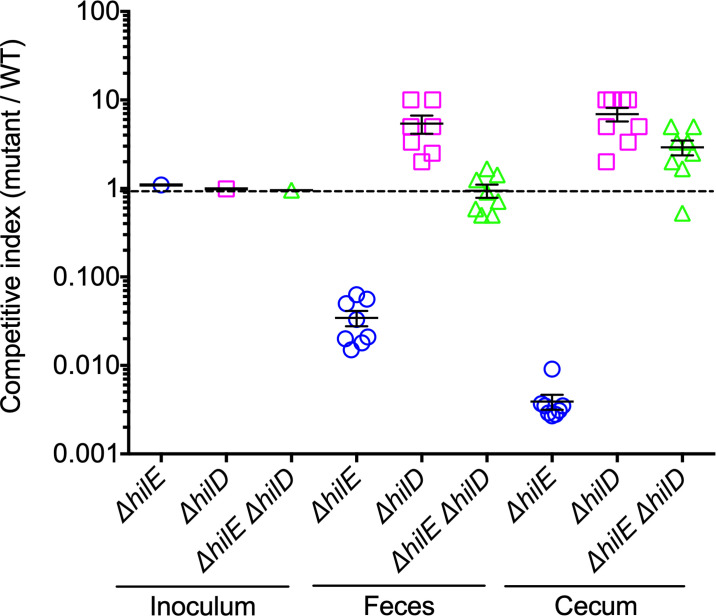
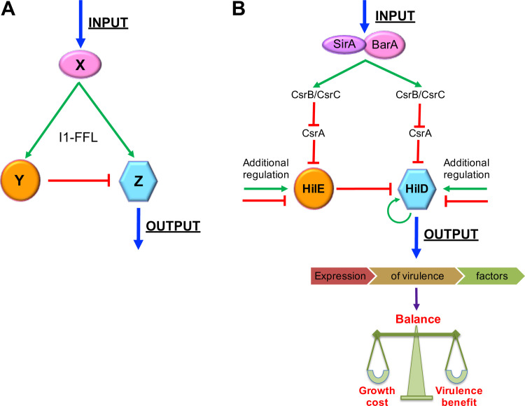
An intricate regulatory network controls the expression of Salmonella virulence genes. The transcriptional regulator HilD plays a central role in this network by controlling the expression of tens of genes mainly required for intestinal colonization. Accordingly, the expression/activity of HilD is highly regulated by multiple factors, such as the SirA/BarA two-component system and the Hcp-like protein HilE. SirA/BarA positively regulates translation of hilD mRNA through a regulatory cascade involving the small RNAs CsrB and CsrC, and the RNA-binding protein CsrA, whereas HilE inhibits HilD activity by protein-protein interaction. In this study, we show that SirA/BarA also positively regulates translation of hilE mRNA through the same mentioned regulatory cascade. Thus, our results reveal a paradoxical regulation exerted by SirA/BarA-Csr on HilD, which involves simultaneous opposite effects, direct positive control and indirect negative control through HilE. This kind of regulation is called an incoherent type-1 feedforward loop (I1-FFL), which is a motif present in certain regulatory networks and represents a complex biological problem to decipher. Interestingly, our results, together with those from a previous study, indicate that HilE, the repressor component of the I1-FFL reported here (I1-FFLSirA/BarA-HilE-HilD), is required to reduce the growth cost imposed by the expression of the genes regulated by HilD. Moreover, we and others found that HilE is necessary for successful intestinal colonization by Salmonella. Thus, these findings support that I1-FFLSirA/BarA-HilE-HilD cooperates to control the precise amount and activity of HilD, for an appropriate balance between the growth cost and the virulence benefit generated by the expression of the genes induced by this regulator. I1-FFLSirA/BarA-HilE-HilD represents a complex regulatory I1-FFL that involves multiple regulators acting at distinct levels of gene expression, as well as showing different connections to the rest of the regulatory network governing Salmonella virulence.


Conflict of interest statement

The authors have declared that no competing interests exist.

Figures

Fig 1
Fig 1. CsrA represses expression of hilE.
(A) HilE-FLAG levels in the WT S. Typhimurium strain carrying a chromosomal FLAG-tagged hilE gene in the absence or presence of pMPM-K3 vector or the pK3-CsrA plasmid, which expresses CsrA from a constitutive promoter, were analyzed by Western blotting using monoclonal anti-FLAG antibodies. As a control for protein loading, the expression of GroEL was also determined using polyclonal anti-GroEL antibodies. A WT S. Typhimurium strain without the FLAG-tagged hilE gene was used as negative control (-). (B) β-galactosidase activity of the translational hilE-lacZ fusion contained in the philE-lacZ plasmid was determined in the WT S. Typhimurium strain in the absence or presence of pMPM-K3 vector or the pK3-CsrA plasmid. Data represent the average of three independent experiments done in duplicate. Error bars denote the standard deviations. Statistically different values are indicated (***, p-value < 0.0001). Western blot and β-galactosidase assays were performed with samples taken from bacterial cultures grown in LB at 37°C.
Fig 2
Fig 2. CsrA specifically interacts with the leader region of the hilE transcript.
(A) CsrA binding to the hilE RNA was analyzed using EMSAs by incubating labeled hilE RNA (0.2 nM) with increasing concentrations of purified CsrA (0, 2.3, 4.7, 9.4, 19, 38, 75, 150 and 300 nM). Positions of bound and free RNA are marked. These experiments were performed three times and a representative gel is shown. (B) RNA competition experiment where labeled hilE RNA (0.2 nM) was combined with 1, 10 and 100 nM of unlabeled specific (hilE) or non-specific (phoB) competitor RNA and incubated with 0 and 75 nM of purified CsrA. Positions of bound and free RNA are marked. A representative gel from two independent assays is shown.
Fig 3
Fig 3. SirA/BarA and CsrB/CsrC positively regulate the expression of hilE.
(A) Western blot analysis of HilE-FLAG expression in the WT S. Typhimurium strain and its derivative ΔsirA, ΔcsrB, ΔcsrC and ΔcsrB ΔcsrC mutants carrying a chromosomal FLAG-tagged hilE gene, using monoclonal anti-FLAG antibodies. As a protein loading control, the expression of GroEL was also determined using polyclonal anti-GroEL antibodies. (B) β-galactosidase activity of the translational hilE-lacZ fusion contained in the philE-lacZ plasmid was determined in the WT S. Typhimurium strain and its derivative ΔbarA, ΔsirA, ΔcsrB, ΔcsrC and ΔcsrB ΔcsrC mutants. (C) β-galactosidase activity of the translational hilE-lacZ fusion contained in the philE-lacZ plasmid was determined in the WT S. Typhimurium strain and its derivative ΔsirA mutant in the absence or presence of pMPM-K3 vector, pK3-SirA or pK3-CsrB plasmids, which express SirA and CsrB, respectively, from a constitutive promoter. Data represent the average of three independent experiments done in duplicate. Error bars symbolize the standard deviations. Statistically different values are indicated (***, p-value < 0.0001). Western blot and β-galactosidase assays were performed with samples taken from bacterial cultures grown in LB at 37°C.
Fig 4
Fig 4. HilE represses the expression of genes regulated by HilD.
(A) CAT activity of the hilA-cat, invF-cat, ssaG-cat, ssrAB-cat, pipB-cat and sopB-cat transcriptional fusions, contained in plasmids philA-cat, pinvF-cat, pssaG-cat, pssrAB-cat, ppipB-cat and psopB-cat, respectively, was determined in the WT S. Typhimurium strain and its derivative ΔhilE, ΔhilD and ΔhilE ΔhilD mutants. (B) CAT activity of the hilA-cat transcriptional fusion, contained in the philA-cat plasmid, was determined in the WT S. Typhimurium strain and its derivative ΔhilE, sirA::Tn10d and ΔhilE sirA::Tn10d mutants in the absence or presence of pMPM-K3 or pMPM-K6 vectors, the pK3-SirA plasmid expressing SirA from a constitutive promoter or the pK6-HilE plasmid expressing HilE from an arabinose-inducible promoter. Expression of HilE from pK6-HilE was induced by adding 0.001% L-arabinose to the medium. (C) CAT activity of the hilA-cat transcriptional fusion, contained in the philA-cat plasmid, was determined in the WT S. Typhimurium strain and its derivative ΔhilD mutant carrying the pMPM-K6 vector or the pK6-HilD plasmid, which expresses HilD from an arabinose-inducible promoter. Expression of HilD from pK6-HilD was induced with 0%, 0.0001% or 0.001% L-arabinose. CAT-specific activity was obtained from samples collected from bacterial cultures grown in LB at 37°C. Data represent the average of three independent experiments done in duplicate. Error bars indicate the standard deviations. Statistically different values are indicated (**, p-value < 0.01; ***, p-value < 0.001; ****, p-value < 0.0001).
Fig 5
Fig 5. HilE negatively controls the bistable expression of SPI-1.
Flow cytometry analysis of GFP fluorescence expression of the invF-gfp transcriptional fusion contained in the low copy pMPMA3ΔPlac PinvF-gfp[LVA]/R plasmid in different S. Typhimurium strains. (A) GFP expression in cultures of the WT strain and its derivative ΔhilE and ΔhilE ΔhilD mutants carrying the invF-gfp fusion, as well as in cultures of the WT strain carrying the gfp reporter gene without a promoter (negative control). (B) GFP expression in cultures of the sirA::Tn10d and ΔhilE sirA::Tn10d mutants carrying the invF-gfp fusion and the pMPM-K4Ω vector or the pK4-SirA plasmid expressing SirA. The percentage of the GFP+ population of each strain is indicated on the graphs. Data shown are representative of two independent experiments performed with ten independent bacterial colonies.
Fig 6
Fig 6. Detrimental effect on bacterial growth by the expression of genes controlled by HilD.
In vitro competitive growth (A) between WT S. Typhimurium strain and isogenic ΔhilE mutant, (B) between WT S. Typhimurium strain and isogenic ΔhilD mutant, (C) between WT S. Typhimurium strain and isogenic ΔhilE ΔhilD mutant, (D) between the ΔsirA and ΔhilE sirA::Tn10d mutants carrying the pMPM-K3 vector, and (E) between the ΔsirA and ΔhilE sirA::Tn10d mutants carrying the pK3-SirA plasmid expressing SirA, was determined by counting CFUs for each strain from samples taken at 0, 2, 4, 6 and 8 h of bacterial mixed cultures grown in LB at 37°C with shaking. CFUs are represented as fraction (percentage) of each strain in the respective mixed culture. Data represent the average of three independent experiments.
Fig 7
Fig 7. HilE is required for the intestinal colonization of mice by S. Typhimurium.
Groups of eight streptomycin-pretreated mice were orally inoculated with a mixed bacterial suspension containing an equal amount of CFUs (0.5 x 10⁶) of the WT S. Typhimurium strain and the respective mutant (WT + ΔhilE, WT + ΔhilD or WT + ΔhilE ΔhilD). At 3 days post-infection, CFUs from feces and cecum were counted for each strain and a Competitive Index (CI; CFUs mutant / CFUs WT) was obtained. A CI was also determined for the respective inoculum. Data for each mouse or inoculum were log-transformed and plotted. CIs are indicated as follows: WT/ΔhilE, blue circles; WT/ΔhilD, pink squares; and WT/ΔhilE ΔhilD, green triangles. Bars denote the standard error of the mean for each experimental group. CI of 1.0, represented by a horizontal dotted line, indicates that the two strains (WT and mutant) were present in equivalent numbers.
Fig 8
Fig 8. Structure and role of I1-FFLSirA/BarA-HilE-HilD in Salmonella virulence.
(A) Structure of the incoherent type-1 feedforward loop (I1-FFL) network motif, where the factor X activates factors Z and Y, and Y represses Z; thus, X exerts opposite controls on Z. (B) Structure and function of I1-FFLSirA/BarA-HilE-HilD. Molecules present in the mammalian intestines, such as short-chain fatty acids, have been shown to activate the SirA/BarA two-component system. SirA/BarA induces expression of both HilD and HilE, through the sRNAs CsrB and CsrC, which bind to CsrA and thus counteract CsrA-mediated repression on the hilD and hilE transcripts. HilE inhibits activity of HilD by protein-protein interaction. Therefore, SirA/BarA simultaneously exerts positive and negative control on HilD, which cooperates for the appropriate balance between the growth cost and the virulence benefit generated by the expression of tens of virulence genes regulated by HilD. Additional positive and negative regulation on HilD and HilE also would control the expression level of the genes regulated by HilD. See text for more details. Green arrows and red blunt-end lines indicate positive and negative control, respectively.

Similar articles

Cited by

References

    1. Haraga A, Ohlson MB, Miller SI. Salmonellae interplay with host cells. Nat Rev Microbiol. 2008;6(1):53–66. doi: 10.1038/nrmicro1788 . - DOI - PubMed
    1. Fàbrega A, Vila J. Salmonella enterica serovar Typhimurium skills to succeed in the host: virulence and regulation. Clin Microbiol Rev. 2013;26(2):308–41. Epub 2013/04/05. doi: 10.1128/CMR.00066-12 . - DOI - PMC - PubMed
    1. Deng W, Marshall NC, Rowland JL, McCoy JM, Worrall LJ, Santos AS, et al.. Assembly, structure, function and regulation of type III secretion systems. Nat Rev Microbiol. 2017;15(6):323–37. Epub 2017/04/11. doi: 10.1038/nrmicro.2017.20 . - DOI - PubMed
    1. Ilyas B, Tsai CN, Coombes BK. Evolution of Salmonella-host cell interactions through a dynamic bacterial genome. Front Cell Infect Microbiol. 2017;7:428. Epub 2017/10/17. doi: 10.3389/fcimb.2017.00428 . - DOI - PMC - PubMed
    1. Dos Santos AMP, Ferrari RG, Conte-Junior CA. Virulence factors in Salmonella Typhimurium: the sagacity of a bacterium. Curr Microbiol. 2019;76(6):762–73. Epub 2018/05/23. doi: 10.1007/s00284-018-1510-4 . - DOI - PubMed

Publication types

MeSH terms