. 2021 May 28;16(5):e0252624.
doi: 10.1371/journal.pone.0252624. eCollection 2021.

Duodenases are a small subfamily of ruminant intestinal serine proteases that have undergone a remarkable diversification in cleavage specificity

Affiliations

Duodenases are a small subfamily of ruminant intestinal serine proteases that have undergone a remarkable diversification in cleavage specificity

Zhirong Fu et al. PLoS One. .

Abstract

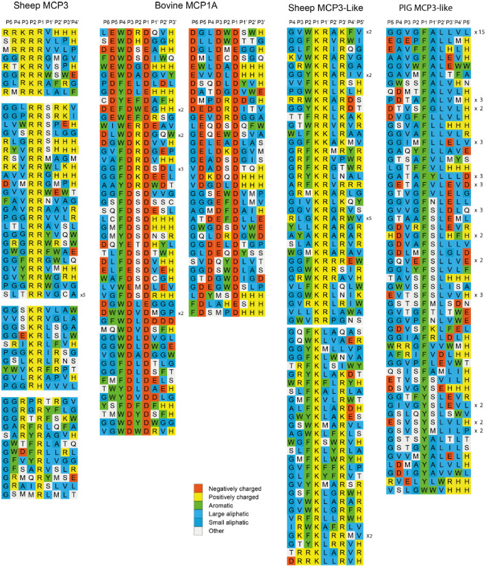
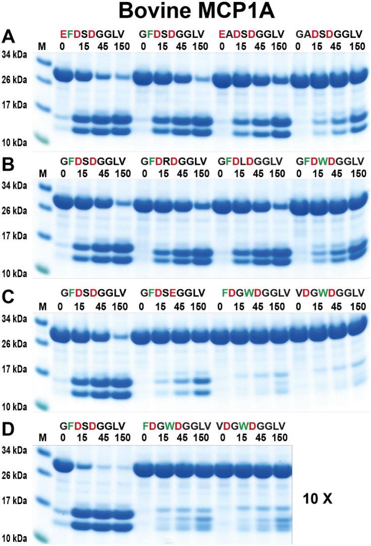
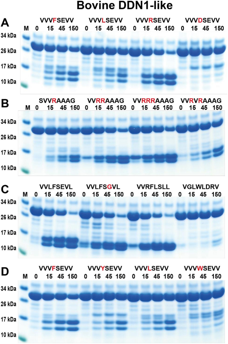
Ruminants have a very complex digestive system adapted for the digestion of cellulose rich food. Gene duplications have been central in the process of adapting their digestive system for this complex food source. One of the new loci involved in food digestion is the lysozyme c locus where cows have ten active such genes compared to a single gene in humans and where four of the bovine copies are expressed in the abomasum, the real stomach. The second locus that has become part of the ruminant digestive system is the chymase locus. The chymase locus encodes several of the major hematopoietic granule proteases. In ruminants, genes within the chymase locus have duplicated and some of them are expressed in the duodenum and are therefore called duodenases. To obtain information on their specificities and functions we produced six recombinant proteolytically active duodenases (three from cows, two from sheep and one from pigs). Two of the sheep duodenases were found to be highly specific tryptases and one of the bovine duodenases was a highly specific asp-ase. The remaining two bovine duodenases were dual enzymes with potent tryptase and chymase activities. In contrast, the pig enzyme was a chymase with no tryptase or asp-ase activity. These results point to a remarkable flexibility in both the primary and extended specificities within a single chromosomal locus that most likely has originated from one or a few genes by several rounds of local gene duplications. Interestingly, using the consensus cleavage site for the bovine asp-ase to screen the entire bovine proteome, it revealed Mucin-5B as one of the potential targets. Using the same strategy for one of the sheep tryptases, this enzyme was found to have potential cleavage sites in two chemokine receptors, CCR3 and 7, suggesting a role for this enzyme to suppress intestinal inflammation.


Conflict of interest statement

The authors have read the journal’s policy and the authors of this manuscript have the following competing interests: LG is a paid employee of GDL Pharmaceutical Consulting and Contracting and JK is a paid employee of Tosoh Bioscience LLC. There are no patents, products in development or marketing products to declare. This does not alter our adherence to PLOS ONE policies on sharing data and materials.

Figures

Fig 1
Fig 1. The lysozyme c locus.
The lysozyme c locus with flanking genes are shown in scale in a panel of different vertebrate species from fish to mammals to highlight the massive expansion of lysozyme c gene that has occurred in ruminants as here represented by sheep and cow.
Fig 2
Fig 2. The chymase locus and a PCR analysis of the expression of duodenases in cow duodenum.
The chymase locus of four mammalian species, human, bovine, sheep and pig is presented in panel A. This is an in-scale presentation of this locus including a few flanking non-lysozyme genes. Panel B shows a PCR analysis of the originally five duodenases in the early version of the cow chymase locus. As can be seen from the figure, only three of the genes show a detectable PCR band of the correct size. In a recent genome update it has been shown that these three genes are complete and most likely functional and the fourth is a pseudogene. This locus does now in the most recent genome update only contain four duodenase genes, three active and one pseudogene. The most recent update of the sheep locus now contains five duodenase genes where four seems to be functional. The second MCP3-like marked with a red dot is most likely non-functional. The genes we have characterized are marked with stars and the ones not yet analysed by red dots. The stars for the bovine enzymes are colour coded as for panel B, the PCR analysis.
Fig 3
Fig 3. A phylogenetic analysis in the form of a phylogenetic tree of chymase locus genes.
A number of mammalian chymase locus genes have been analysed for sequence relatedness using the neighbour joining algorithm. Human complement factor B and a few coagulation factor X proteins were used as outgroup. Duodenases form a clear subfamily distinct from the cathepsin G, granzymes and chymases. The six genes analysed in more detail for their cleavage specificity is marked by stars as in the Fig 2 and the genes not yet characterized are marked with a red dot, also similar to the Fig 2.
Fig 4
Fig 4. SDS-PAGE of the recombinant sheep, cow and pig duodenases.
The recombinant sheep MCP3 was expressed in a baculovirus expression system. These proenzymes were first purified on Ni-NTA beads (-EK) and then activated by removal of the His6-tag by enterokinase digestion (+EK). Following activation, the enzyme was further purified over heparin-Sepharose columns. In the right part of the figure we show the recombinant cow, pig and sheep duodenases, cow DDN1, cow DDN-like, cow MCP1A, Pig MCP3-like and the second sheep duodenase sheep MCP3-like. All these proteases were produced in the human cell line HEK293-EBNA using the episomal vector pCEP-Pu2. After purification on IMAC columns they were activated by enterokinase cleavage. The proteins are shown before and after enterokinase cleavage. Enterokinase removes the N terminal six his tag and the enterokinase site resulting in a reduction in size of the protein by approximately 1.5 kDa. The enzymes were after purification analysed by separation on SDS-PAGE and visualized with Coomassie Brilliant Blue staining.
Fig 5
Fig 5. Chromogenic substrate analysis of the sheep, cow and pig duodenases.
A panel of chromogenic substrates was used to determine the primary specificity of the different duodenases. Specificities of the human chymase and human thrombin are presented as reference. The panel includes different chymase, elastase, tryptase and asp-ase substrates. The amino acid sequences of the substrates are listed at the left side of the panels. The analyses were done in triplicates and the standard deviation bars within these triplicates is presented in the figure.
Fig 6
Fig 6. Phage display analysis of sheep MCP3, cow MCP1A, sheep MCP3-like and pig MCP3-like.
After the last selection step of the phage display analysis, phages released by proteolytic cleavage of the three proteases were isolated and the sequences encoding the nonamers were determined. The general sequence of the T7 phage capsid proteins are PGG(X)9HHHHHH, where (X)9 indicates the randomized nonamers. The protein sequences were aligned into a P4-P3´ consensus, where cleavage occurs between positions P1 and P1´. If the sequence as found more than once this is indicated by the corresponding number to the left of the sequence. The amino acids are colour coded according to the side chain properties as indicated in the legend.
Fig 7
Fig 7. Verification of the cleavage specificity of sheep MCP3 using recombinant protein substrates.
Panel A shows the overall structure of the recombinant protein substrates used for analysis of the efficiency in cleavage by the enzyme. In these substrates two thioredoxin molecules are positioned in tandem and the proteins have a His6-tag positioned in their C termini. The different cleavable sequences are inserted in the linker region between the two thioredoxin molecules using two unique restriction sites, one Bam HI and one SalI site, which are indicated in the bottom of panel A. In panel B, a schematic representation of a cleavage reaction is presented. In Panels C to G, the cleavage of several substrates by sheep MCP3 is presented. The sequences of the different substrates are indicated above the pictures of the gels. The time of cleavage in minutes is also indicated above the corresponding lanes of the different gels. The uncleaved substrates have a molecular weight of ~25 kDa and the cleaved substrates appear as two closely located bands with a size of 12–13 kDa. Residues of particular interest and that may differ between different substrates are marked in red or green for an easy identification.
Fig 8
Fig 8. Verification of the cleavage specificity of bovine MCP1A by the use of recombinant protein substrates.
Panels from A to D show the cleavage of a number of substrates by bovine MCP1A. The sequences of the different substrates are indicated above the pictures of the gels. The time of cleavage in minutes is also indicated above the corresponding lanes of the different gels. The uncleaved substrates have a molecular weight of ~25 kDa and the cleaved substrates appear as two closely located bands with a size of 12–13 kDa. Residues of particular interest and that may differ between different substrates are marked in red or green for an easy identification. In panel D, we used 10-fold more enzyme compared to the other cleavage reactions.
Fig 9
Fig 9. Verification of the cleavage specificity of sheep MCP3-like using recombinant protein substrates.
Panels A to E show the cleavage of several substrates by MCP3-like. The sequences of the different substrates are indicated above the pictures of the gels. The time of cleavage in minutes is also indicated above the corresponding lanes of the different gels. The uncleaved substrates have a molecular weight of ~25 kDa and the cleaved substrates appear as two closely located bands with a size of 12–13 kDa. Residues of particular interest and that may differ between different substrates are marked in red or green for an easy identification.
Fig 10
Fig 10. Verification of the cleavage specificity of pig MCP3-like using recombinant protein substrates.
Panels A to D shows the cleavage of several substrates by the pig MCP3-like. The sequence of the different substrates are indicated above the pictures of the gels. The time of cleavage in minutes is also indicated above the corresponding lanes of the different gels. The uncleaved substrates have a molecular weight of ~25 kDa and the cleaved substrates appear as two closely located bands with a size of 12–13 kDa. Residues of particular interest and that may differ between different substrates are marked in red for an easy identification.
Fig 11
Fig 11. Analysis of the cleavage specificity of bovine duodenase DDN1 using recombinant protein substrates.
Panels A to D shows the cleavage of several substrates by the bovine DDN1. The sequence of the different substrates are indicated above the pictures of the gels. The time of cleavage in minutes is also indicated above the corresponding lanes of the different gels. The uncleaved substrates have a molecular weight of ~25 kDa and the cleaved substrates appear as two closely located bands with a size of 12–13 kDa. Residues of particular interest and that may differ between different substrates are marked in red or green for an easy identification.
Fig 12
Fig 12. Analysis of the cleavage specificity of bovine duodenase DDN1-like using recombinant protein substrates.
Panels A to D shows the cleavage of several substrates by the bovine DDN-like. The sequence of the different substrates are indicated above the pictures of the gels. The time of cleavage in minutes is also indicated above the corresponding lanes of the different gels. The uncleaved substrates have a molecular weight of ~25 kDa and the cleaved substrates appear as two closely located bands with a size of 12–13 kDa. Residues of particular interest and that may differ between different substrates are marked in red for an easy identification.
Fig 13
Fig 13. Schematic representation of three potential targets for bovine MCP1A and sheep MCP3-like.
In panel A, we show a schematic picture of bovine Mucin 5B with the different regions of the protein marked in different colours and where the six consensus sites for bovine MCP1A are marked by red arrows. The sequence of the six potential cleavage sites are listed below the schematic figure. In panel B, we show the schematic structure of two chemokine receptors having potential cleavage site for sheep MCP3-like. The position of the membrane is marked with a black line and the potential cleavage sites by red arrows. The sequences of the potential cleavage sites are listed below the schematic figure.
Fig 14
Fig 14. Sequence alignment of a panel of cow, sheep and pig duodenases.
The figure shows the sequence alignment where three residues of importance for target selectivity are highlighted, residues 189, 216 and 226 (chymotrypsin numbering). These three residues form the catalytic pocket and as can be seen cow MCP1A differ from the other duodenases in position 226 where it has an Arg, which is marked by a red circle. This position is of major importance for its different primary specificity as being an asp-ase. Sheep DDN1-like lacks the N terminal part of the mature protease and is most likely a pseudogene.

Similar articles

Cited by

References

    1. Cross M, Mangelsdorf I, Wedel A, Renkawitz R. Mouse lysozyme M gene: isolation, characterization, and expression studies. Proc Natl Acad Sci U S A. 1988;85(17):6232–6. Epub 1988/09/01. 10.1073/pnas.85.17.6232 . - DOI - PMC - PubMed
    1. Irwin DM. Genomic organization and evolution of ruminant lysozyme c genes. Dongwuxue Yanjiu. 2015;36(1):1–17. Epub 2015/03/03. 10.13918/j.issn.2095-8137.2015.1.1 . - DOI - PMC - PubMed
    1. Irwin DM. Evolution of cow nonstomach lysozyme genes. Genome/National Research Council Canada = Genome/Conseil national de recherches Canada. 2004;47(6):1082–90. 10.1139/g04-075 . - DOI - PubMed
    1. Henke M, Hobom G, Senft B, Seyfert HM. Structural deviations in a bovine low expression lysozyme-encoding gene active in tissues other than stomach. Gene. 1996;178(1–2):131–7. Epub 1996/10/31. 10.1016/0378-1119(96)00352-6 . - DOI - PubMed
    1. Litwinczuk Z, Krol J, Brodziak A, Barlowska J. Changes of protein content and its fractions in bovine milk from different breeds subject to somatic cell count. J Dairy Sci. 2011;94(2):684–91. Epub 2011/01/25. 10.3168/jds.2010-3217 . - DOI - PubMed

Publication types

LinkOut - more resources