. 2022 Jun 22;118(7):1835-1848.
doi: 10.1093/cvr/cvab183.

Vascular histopathology and connective tissue ultrastructure in spontaneous coronary artery dissection: pathophysiological and clinical implications

Affiliations

Vascular histopathology and connective tissue ultrastructure in spontaneous coronary artery dissection: pathophysiological and clinical implications

Marios Margaritis et al. Cardiovasc Res. .

Erratum in

Abstract

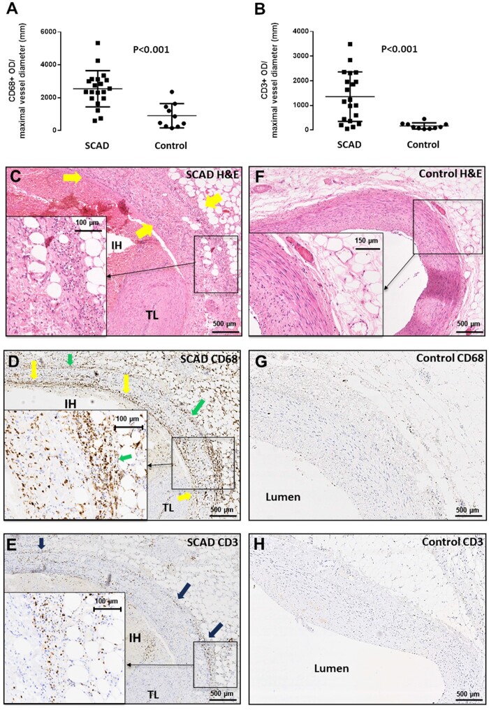
Aims: Spontaneous coronary artery dissection (SCAD) is a cause of acute coronary syndromes and in rare cases sudden cardiac death (SCD). Connective tissue abnormalities, coronary inflammation, increased coronary vasa vasorum (VV) density, and coronary fibromuscular dysplasia have all been implicated in the pathophysiology of SCAD but have not previously been systematically assessed. We designed a study to investigate the coronary histological and dermal collagen ultrastructural findings in SCAD.

Methods and results: Thirty-six autopsy SCAD cases were compared with 359 SCAD survivors. Coronary and myocardial histology and immunohistochemistry were undertaken. Transmission electron microscopy (TEM) of dermal extracellular matrix (ECM) components of n = 31 SCAD survivors and n = 16 healthy volunteers were compared. Autopsy cases were more likely male (19% vs. 5%; P = 0.0004) with greater proximal left coronary involvement (56% vs. 18%; P < 0.0001) compared to SCAD survivors. N = 24 (66%) of cases showed no myocardial infarction on macro- or microscopic examination consistent with arrhythmogenic death. There was significantly (P < 0.001) higher inflammation in cases with delayed-onset death vs. sudden death and significantly more inflammation surrounding the dissected vs. non-dissected vessel segments. N = 17 (47%) cases showed limited intimal fibro-elastic thickening but no features of fibromuscular dysplasia and no endothelial or internal elastic lamina abnormalities. There were no differences in VV density between SCAD and control cases. TEM revealed no general ultrastructural differences in ECM components or markers of fibroblast metabolic activity.

Conclusions: Assessment of SCD requires careful exclusion of SCAD, particularly in cases without myocardial necrosis. Peri-coronary inflammation in SCAD is distinct from vasculitides and likely a reaction to, rather than a cause for SCAD. Coronary fibromuscular dysplasia or increased VV density does not appear pathophysiologically important. Dermal connective tissue changes are not common in SCAD survivors.

Keywords: Autopsy; Collagen; Electron microscopy; Haematoma; Inflammation; Spontaneous coronary artery dissection; Sudden cardiac death; Vascular.


Figures

Graphical Abstract
Graphical Abstract
Figure 1
Figure 1
Composition of peri-adventitial inflammatory cell infiltrate in SCAD. Compared to age- and sex-matched control cases (n = 10), SCAD autopsy cases (n = 20) showed significantly higher infiltration with CD68+ macrophages (A, P < 0.001) and CD3+ T cells (B, P < 0.001). This infiltrate comprising lymphocytes, macrophages, and eosinophils was visualized in H&E staining (C) and spatially analysed with IHC. CD68+ cells were abundant throughout (D), whereas CD3+ T cells were less numerous and localized to the adventitial border (E). Control sections did not feature these findings (F–H). All comparisons between groups were made using unpaired t-test on log-transformed values.
Figure 2
Figure 2
Association between the degree of peri-adventitial inflammatory infiltrate and time interval from SCAD symptom onset to death. Increased degree of peri-adventitial inflammatory cell infiltration was significantly associated with increased time from symptom onset to death (A, n = 27, P = 0.006, χ test). CD68+ and CD3+ staining showing a higher macrophage (B, CD68+, P < 0.001, n = 17), and T-cell infiltrate (B, CD3+, P = 0.03, n = 17) in cases with more than 24 h delay between onset of SCAD-related symptoms and death. Comparisons made using an unpaired t-test on log-transformed values. Panels (C–I) provide examples of autopsy cases belonging to the delayed-onset (C–F) vs. rapid-onset (G–I) death groups.
Figure 3
Figure 3
Association between the degree of peri-adventitial inflammatory infiltrate and proximity to dissected portion of the medial layer. In SCAD cases, we observed significantly higher inflammatory area (A, P < 0.0001; n = 18) and denser peri-adventitial inflammatory cell infiltrate (B, P < 0.0001, n = 18) adjacent to dissected segments vs. non-dissected coronary segments. In a typical SCAD section (C), there is denser reactive adventitial tissue (green arrows) surrounding the intramural haematoma (IH) vs. areas adjacent to healthy, non-dissected portions of the medial layer (black arrows). Similarly, IHC showed that the number of CD68+ macrophages (D, P < 0.0001, n = 8) and CD3+ T-lymphocytes (E, P = 0.016, n = 8) was higher in the adventitia surrounding the dissected vs. non-dissected coronary circumference. All comparisons between dissected and non-dissected segments were made using paired t-test.
Figure 4
Figure 4
VV density in SCAD. VV in SCAD coronary sections (n = 20) vs. control cases (n = 10) (A, P = 0.47) or total CD31+ optical density in the vascular media adjusted for maximal vessel diameter (B, P = 0.43). Panels (C and D) are representative SCAD and control microphotographs of CD31-stained sections, respectively. VV density comparing rapid-onset (<24 h from symptom to death) to delayed-onset (>24 h) SCAD fatalities (E and F, G and H representative microphotographs for <24 (n = 6) and >24 h (n = 8) groups, respectively. P = NS). All comparisons between groups were made using an unpaired t-test on log-transformed values.
Figure 5
Figure 5
Intramural haematoma features from the SCAD autopsy case series. (A and B) Sequential H&E sections from the same autopsy case. Distally only a small proportion of the total vessel circumference is affected (A), leaving the true lumen (TL) relatively patent, whereas proximally the false lumen envelops almost the entire vessel causing significant luminal compression (B). (C) Intramural haematoma (purple arrow) displaying dense red clot with minimal fibrin formation. (D) Intramural haematoma (purple arrows) showing varying degrees of maturation with fibrin formation, with almost distinct ‘compartmentalization’ of segments within. Yellow arrows: intramural haematoma; TL, true lumen.
Figure 6
Figure 6
Intimal and medial layer features in SCAD. (A) H&E staining of SCAD lesion with medial dissection and intramural haematoma (IH) leading to compression true lumen (TL) compression (black arrows). Moderate fibro-elastic thickening of the endothelial layer (red arrows) but no abnormalities in structure or orientation of medial smooth muscle cells (yellow arrows). (B) EVG staining. Fibro-elastic intimal thickening is well-delineated (red arrows); the IEL clearly visualized and smooth muscle cell medial layer distinguished from intima and adventitia (yellow arrows). (C) CD31 staining shows mature endothelial cells on the intimal surface (green arrows). (D) αSMA showing normal pattern of staining in the non-dissected segment of the media (blue arrows).
Figure 7
Figure 7
Ultrastructural analysis of the main ECM components in SCAD patients vs. HV. Elastin diameter (A), collagen fibril minimum diameter (B) and irregular collagen fibril diameter (C) distributions in SCAD (n = 31) vs. HV (n = 16). Representative images of elastin (D), collagen fibrils (E), and irregular fibrils (F and G). T-test between the averages, standard deviations, minimum and maximum values, and ranges of these parameters was performed between SCAD and HV. No significant differences were observed.

Comment in

References

    1. Adlam D, Alfonso F, Maas A, Vrints C; Writing Committee . European Society of Cardiology, acute cardiovascular care association, SCAD study group: a position paper on spontaneous coronary artery dissection. Eur Heart J 2018;39:3353–3368. - PMC - PubMed
    1. Hayes SN, Kim ESH, Saw J, Adlam D, Arslanian-Engoren C, Economy KE, Ganesh SK, Gulati R, Lindsay ME, Mieres JH, Naderi S, Shah S, Thaler DE, Tweet MS, Wood MJ; American Heart Association Council on Peripheral Vascular Disease; Council on Clinical Cardiology; Council on Cardiovascular and Stroke Nursing; Council on Genomic and Precision Medicine; and Stroke Council . Spontaneous coronary artery dissection: current state of the science: a scientific statement from the American Heart Association. Circulation 2018;137:e523–e557. - PMC - PubMed
    1. Desai S, Sheppard MN.. Sudden cardiac death: look closely at the coronaries for spontaneous dissection which can be missed. A study of 9 cases. Am J Forensic Med Pathol 2012;33:26–29. - PubMed
    1. Vrints CJ. Spontaneous coronary artery dissection. Heart 2010;96:801–808. - PubMed
    1. Kwon TG, Gulati R, Matsuzawa Y, Aoki T, Guddeti RR, Herrmann J, Lennon RJ, Ritman EL, Lerman LO, Lerman A.. Proliferation of coronary adventitial vasa vasorum in patients with spontaneous coronary artery dissection. JACC Cardiovasc Imaging 2016;9:891–892. - PubMed

Publication types

Supplementary concepts