

Molecular remodeling of Cx43, but not structural remodeling, promotes arrhythmias in an arrhythmogenic canine model of nonischemic heart failure
- PMID: 34048725
- PMCID: PMC8963384
- DOI: 10.1016/j.yjmcc.2021.05.012
Molecular remodeling of Cx43, but not structural remodeling, promotes arrhythmias in an arrhythmogenic canine model of nonischemic heart failure
Abstract
Background: Both gap junctional remodeling and interstitial fibrosis have been linked to impaired electrical conduction velocity (CV) and fatal ventricular arrhythmias in nonischemic heart failure (HF). However, the arrhythmogenic role of the ventricular gap junctional Cx43 in nonischemic HF remains in debate. Here, we assessed this in a newly developed arrhythmogenic canine model of nonischemic HF.
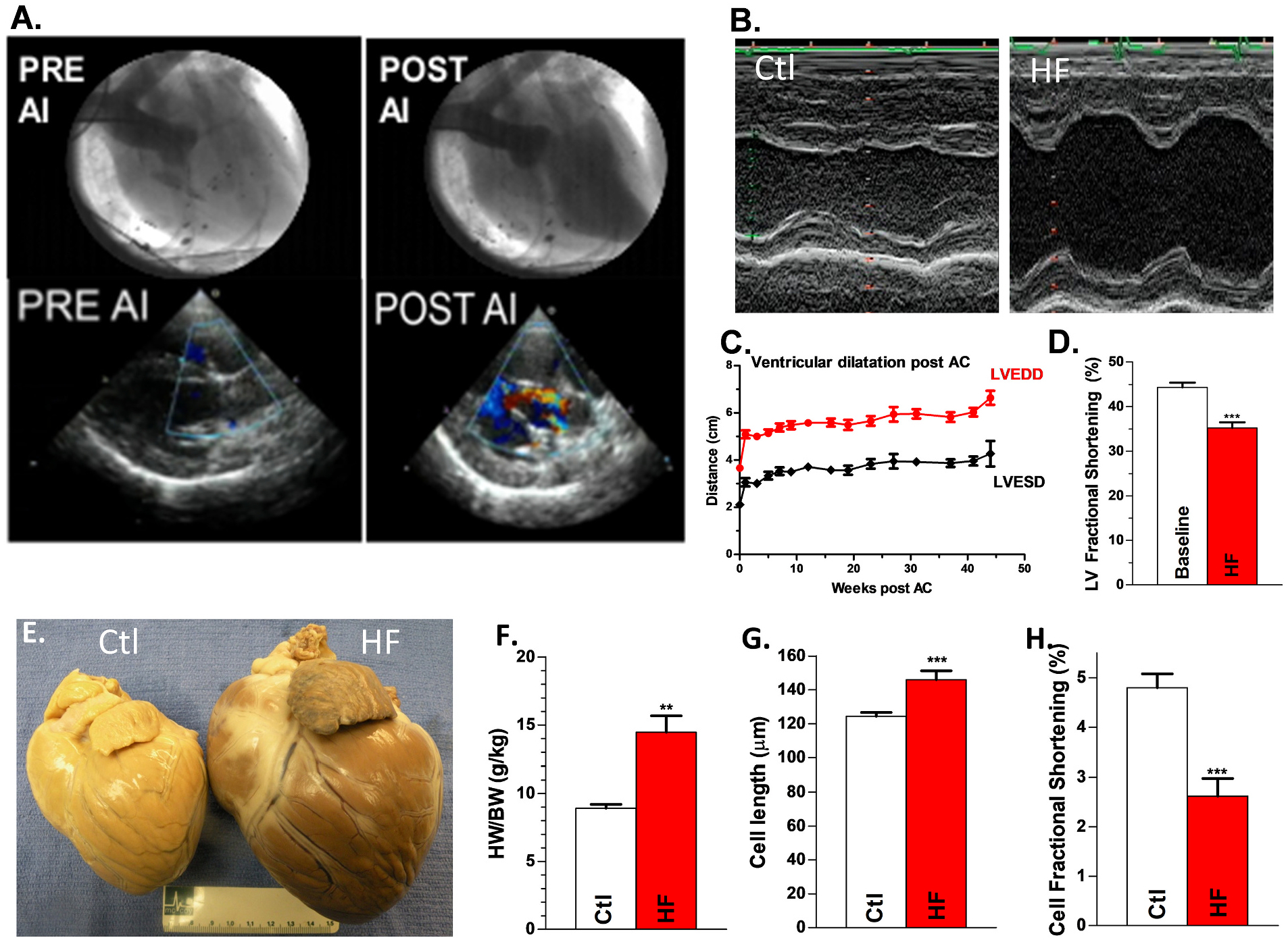
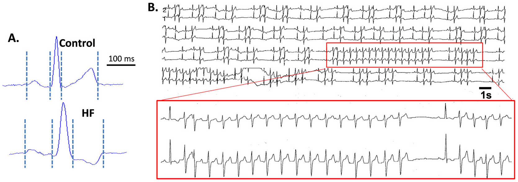
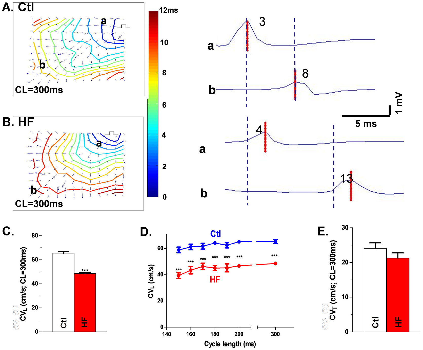
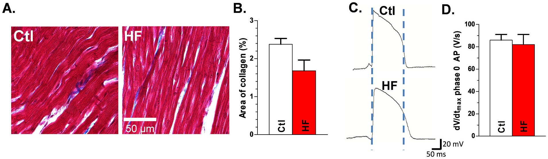
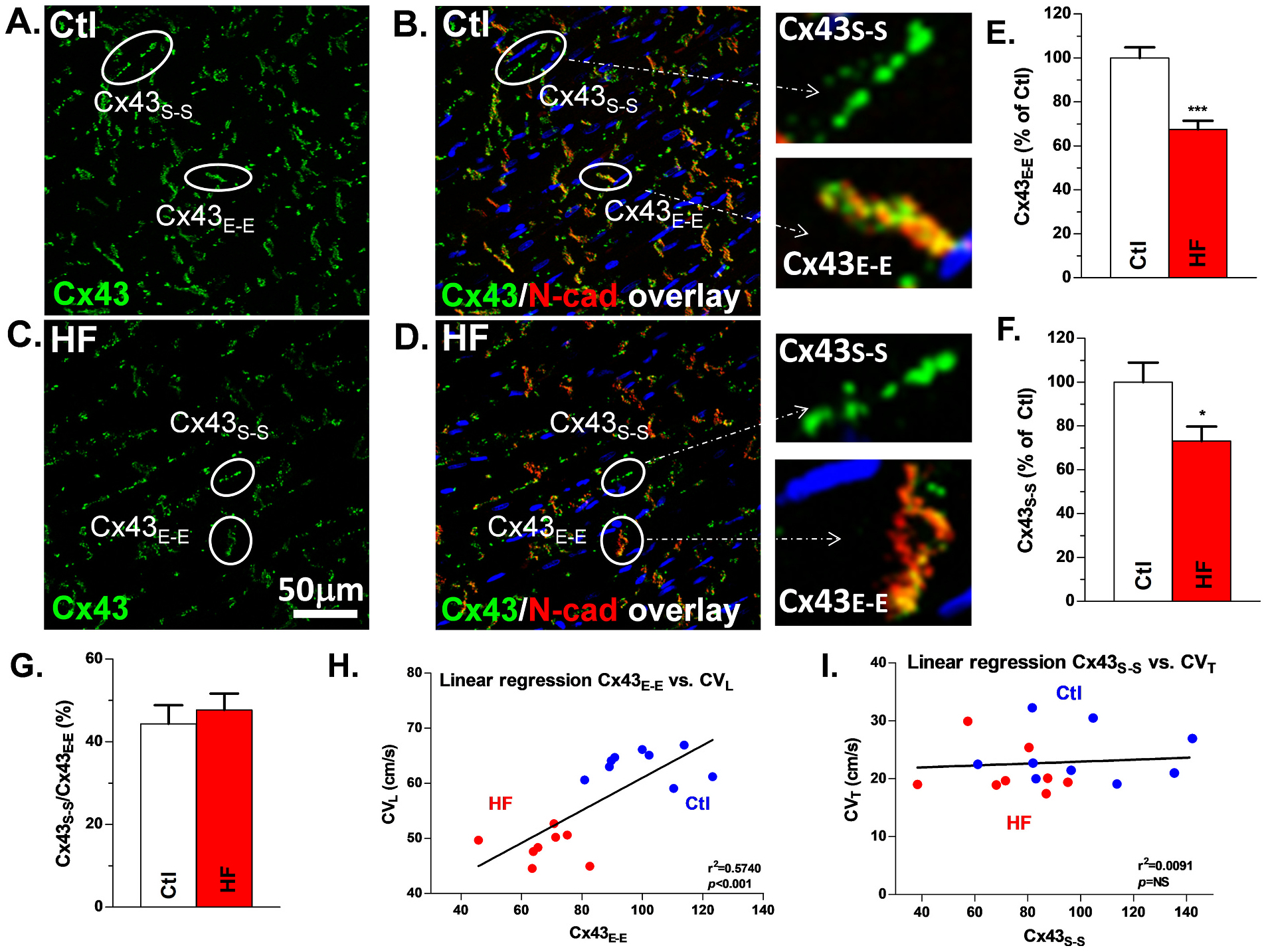
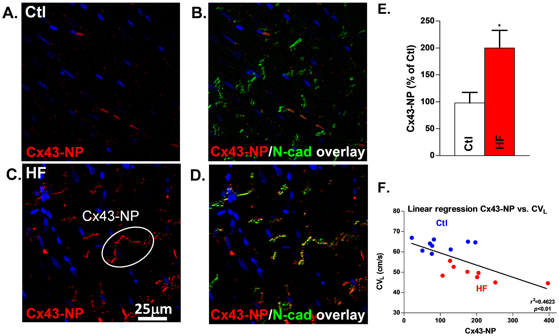
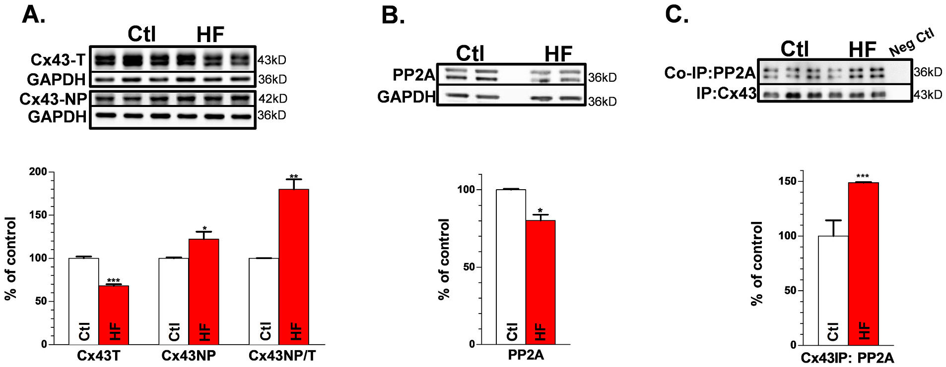
Methods and results: Nonischemic HF was induced in canines by combined aortic valve insufficiency and aortic constriction. Left ventricular (LV) myocardium from HF dogs showed similar pathological changes to that of humans. HF dogs had reduced LV function, widened QRS complexes, and spontaneous nonsustained ventricular tachycardia. CV was measured in intact LV epicardium with high-density grid mapping. Total (Cx43-T) and nonphosphorylated Cx43 (Cx43-NP) and histological interstitial fibrosis were assessed from these mapped LV tissues. Longitudinal CV, which was slowed in HF (49 ± 1 vs. 65 ± 2 cm/s in Ctl), was positively correlated with reduced total junctional Cx43 and negatively correlated with markedly increased junctional Cx43-NP (2-fold) in HF. Cx43 dephosphorylation in HF was associated with enhanced colocalization of PP2A at the level of Cx43. Unchanged action potential upstroke and transverse CV were associated with unaltered Cx43 lateralization and interstitial fibrosis in the nonischemic HF canine LV.
Conclusion: Our unique arrhythmogenic canine model of HF resembles human nonischemic HF (prior to the end stage). Cx43 remodeling occurs prior to the structural remodeling (with lack of fibrosis) in HF and it is crucial in slowed CV and ventricular arrhythmia development. Our findings suggest that altered Cx43 alone is arrhythmogenic and modulation of Cx43 has the anti-arrhythmic therapeutic potential for HF patients.
Keywords: Conduction velocity; Dephosphorylation; Fibrosis; Heart failure; connexin43 remodeling.
Copyright © 2021. Published by Elsevier Ltd.
Conflict of interest statement
Declaration of Competing Interest
None.
Figures
Similar articles
-
Connexin 43 downregulation and dephosphorylation in nonischemic heart failure is associated with enhanced colocalized protein phosphatase type 2A.Circ Res. 2005 Jan 7;96(1):54-63. doi: 10.1161/01.RES.0000152325.07495.5a. Epub 2004 Dec 2. Circ Res. 2005. PMID: 15576650
-
Dynamic changes in conduction velocity and gap junction properties during development of pacing-induced heart failure.Am J Physiol Heart Circ Physiol. 2007 Aug;293(2):H1223-30. doi: 10.1152/ajpheart.00079.2007. Epub 2007 Apr 13. Am J Physiol Heart Circ Physiol. 2007. PMID: 17434978
-
Mechanisms underlying conduction slowing and arrhythmogenesis in nonischemic dilated cardiomyopathy.Circ Res. 2004 Oct 1;95(7):717-25. doi: 10.1161/01.RES.0000144125.61927.1c. Epub 2004 Sep 2. Circ Res. 2004. PMID: 15345654
-
Myocardial gap junctions: targets for novel approaches in the prevention of life-threatening cardiac arrhythmias.Physiol Res. 2008;57 Suppl 2:S1-S13. doi: 10.33549/physiolres.931546. Epub 2008 Mar 28. Physiol Res. 2008. PMID: 18373398 Review.
-
Heart failure as a substrate and trigger for ventricular tachycardia.J Interv Card Electrophysiol. 2019 Dec;56(3):229-247. doi: 10.1007/s10840-019-00623-x. Epub 2019 Oct 9. J Interv Card Electrophysiol. 2019. PMID: 31598875 Review.
Cited by
-
Circadian Regulation of Cardiac Arrhythmias and Electrophysiology.Circ Res. 2024 Mar 15;134(6):659-674. doi: 10.1161/CIRCRESAHA.123.323513. Epub 2024 Mar 14. Circ Res. 2024. PMID: 38484028 Free PMC article. Review.
-
Lethal Arrhythmogenic Role of Left Ventricular Myocardial Interstitial Fibrosis in Apolipoprotein E/Low-Density Lipoprotein Receptor Double-Knockout Mice with Metabolic Dysfunction-Associated Steatohepatitis.Int J Mol Sci. 2024 Dec 27;26(1):144. doi: 10.3390/ijms26010144. Int J Mol Sci. 2024. PMID: 39796002 Free PMC article.
-
Biomarkers of Atrial Fibrillation Recurrence in Patients with Paroxysmal or Persistent Atrial Fibrillation Following External Direct Current Electrical Cardioversion.Biomedicines. 2023 May 16;11(5):1452. doi: 10.3390/biomedicines11051452. Biomedicines. 2023. PMID: 37239123 Free PMC article. Review.
-
Reversible complete left bundle branch block and a wide QRS complex following administration of sodium-glucose cotransporter-2 inhibitor and volume reduction in a patient with systolic heart failure: a case report.Eur Heart J Case Rep. 2024 Sep 14;8(9):ytae512. doi: 10.1093/ehjcr/ytae512. eCollection 2024 Sep. Eur Heart J Case Rep. 2024. PMID: 39345957 Free PMC article.
-
Role of Connexin 43 phosphorylation on Serine-368 by PKC in cardiac function and disease.Front Cardiovasc Med. 2023 Jan 12;9:1080131. doi: 10.3389/fcvm.2022.1080131. eCollection 2022. Front Cardiovasc Med. 2023. PMID: 36712244 Free PMC article. Review.
References
-
- Packer M, Sudden unexpected death in patients with congestive heart failure: a second frontier, Circulation 72 (1985) 681–685. - PubMed
-
- Pogwizd SM, Nonreentrant mechanisms underlying spontaneous ventricular arrhythmias in a model of nonischemic heart failure in rabbits, Circulation 92 (1995) 1034–1048. - PubMed
-
- Pogwizd SM, McKenzie JP, Cain ME, Mechanisms underlying spontaneous and induced ventricular arrhythmias in patients with idiopathic dilated cardiomyopathy, Circulation 98 (1998) 2404–2414. - PubMed
-
- Pogwizd SM, Schlotthauer K, Li L, Yuan W, Bers DM, Arrhythmogenesis and contractile dysfunction in heart failure: roles of sodium-calcium exchange, inward rectifier potassium current, and residual beta-adrenergic responsiveness, Circ. Res 88 (2001) 1159–1167. - PubMed
Publication types
MeSH terms
Substances
Grants and funding
LinkOut - more resources
Full Text Sources
Other Literature Sources
Medical
Research Materials
Miscellaneous
