Comparative Study
. 2021 Jun:68:103403.
doi: 10.1016/j.ebiom.2021.103403. Epub 2021 May 25.

Comparing infectivity and virulence of emerging SARS-CoV-2 variants in Syrian hamsters

Affiliations
Comparative Study

Comparing infectivity and virulence of emerging SARS-CoV-2 variants in Syrian hamsters

Rana Abdelnabi et al. EBioMedicine. 2021 Jun.

Abstract

Background: Within one year after its emergence, more than 108 million people acquired SARS-CoV-2 and almost 2·4 million succumbed to COVID-19. New SARS-CoV-2 variants of concern (VoC) are emerging all over the world, with the threat of being more readily transmitted, being more virulent, or escaping naturally acquired and vaccine-induced immunity. At least three major prototypic VoC have been identified, i.e. the United Kingdom, UK (B.1.1.7), South African (B.1.351) and Brazilian (B.1.1.28.1) variants. These are replacing formerly dominant strains and sparking new COVID-19 epidemics.

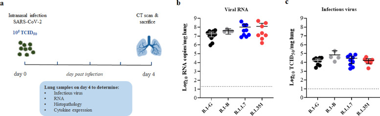
Methods: We studied the effect of infection with prototypic VoC from both B.1.1.7 and B.1.351 variants in female Syrian golden hamsters to assess their relative infectivity and virulence in direct comparison to two basal SARS-CoV-2 strains isolated in early 2020.

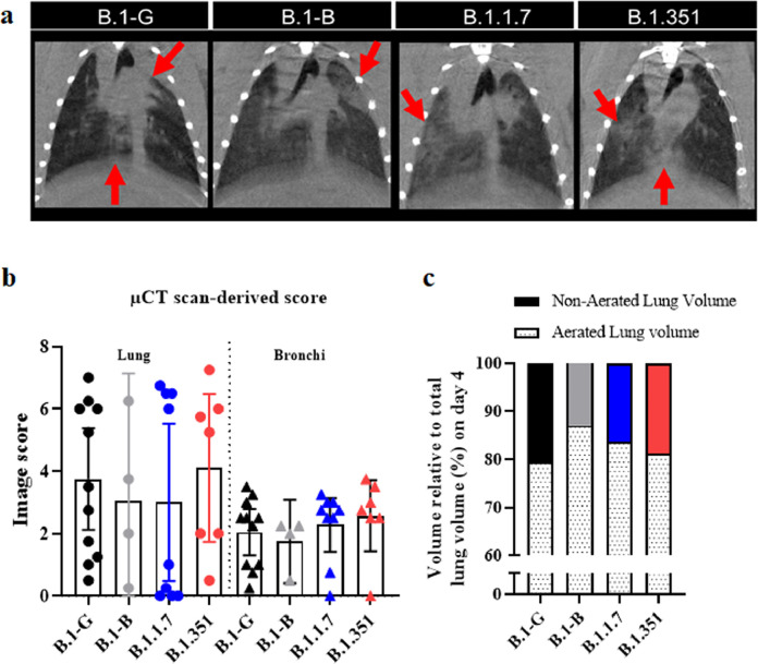
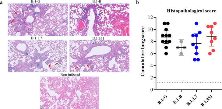
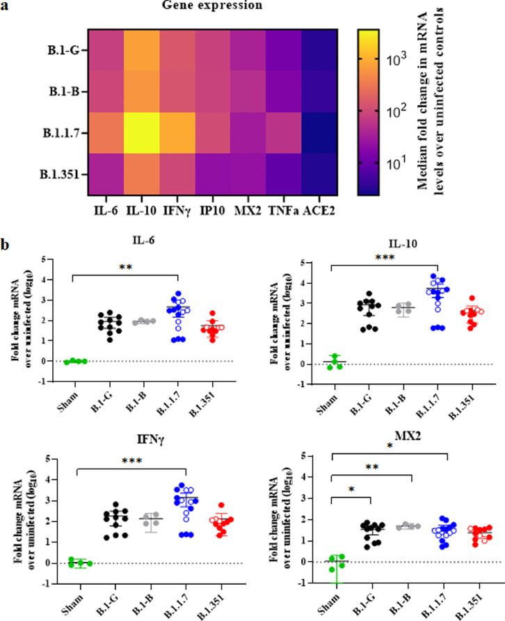
Findings: A very efficient infection of the lower respiratory tract of hamsters by these VoC is observed. In line with clinical evidence from patients infected with these VoC, no major differences in disease outcome were observed as compared to the original strains as was quantified by (i) histological scoring, (ii) micro-computed tomography, and (iii) analysis of the expression profiles of selected antiviral and pro-inflammatory cytokine genes. Noteworthy however, in hamsters infected with VoC B.1.1.7, a particularly strong elevation of proinflammatory cytokines was detected.

Interpretation: We established relevant preclinical infection models that will be pivotal to assess the efficacy of current and future vaccine(s) (candidates) as well as therapeutics (small molecules and antibodies) against two important SARS-CoV-2 VoC.

Funding: Stated in the acknowledgment.

Keywords: Emergence; Hamster model; SARS-CoV-2; Variants of concern (VoC).


Conflict of interest statement

Declaration of Competing Interest Authors declare no conflict of interests.

Figures

Fig 1
Fig. 1
Viral loads from the lungs of Syrian hamsters infected with different SARS-CoV-2 variants. (a) Set-up of the Syrian hamster infection study. (b) Viral RNA levels in the lungs of hamsters infected with 105 TCID50 of B.1-G (n = 11), B.1-B (n = 4), B.1.1.7 (n = 9) or B.1.351 (n = 8) SARS-CoV-2 variants on day 4 post-infection (pi) are expressed as log10 SARS-CoV-2 RNA copies per mg lung tissue. Individual data and mean values with 95% CI are presented. (c) Infectious viral loads in the lungs of hamsters infected with the different SARS-CoV-2 variants at day 4 pi are expressed as log10 TCID50 per mg lung tissue. Individual data and mean values with 95% CI are presented. All data are from at least two independent experiments except for the B.1-B group.
Fig 2
Fig. 2
Micro-CT analysis of lung disease burden in Syrian hamsters infected with different SARS-CoV-2 variants. (a) Representative coronal lung micro-CT images of hamsters infected with B.1-G, B.1-B, B.1.1.7 or B.1.351 SARS-CoV-2 variants at day 4 pi. Red arrows indicate examples of pulmonary infiltrates. (b) Quantification of the micro-CT-derived lung and bronchi disease scores in hamsters infected with 105 TCID50 of B.1-G (n = 10), B.1-B (n = 4), B.1.1.7 (n = 9) or B.1.351 (n = 7) SARS-CoV-2 variants on day 4 post-infection. Individual data per hamster are shown, bars represent mean values with 95% CI. (c) Micro-CT-derived non-aerated lung volume (reflecting the tissue lesion volume) and aerated lung volume relative to total lung volume of hamsters infected with the different SARS-CoV-2 variants. All data are from at least two independent experiments except for the B.1-B group. (For interpretation of the references to color in this figure legend, the reader is referred to the web version of this article.)
Fig 3
Fig. 3
Histopathology of lungs of Syrian hamsters infected with different SARS-CoV-2 variants. (a) Representative H&E images of lungs of hamsters infected with 105 TCID50 of B.1-G (n = 11), B.1-B (n = 4), B.1.1.7 (n = 9) or B.1.351 (n = 8) SARS-CoV-2 variants at day 4 pi. The lungs of hamsters infected with any of the SARS-CoV-2 variants show peri-bronchial inflammation (blue arrows) and bronchopneumonia in the surrounding alveoli (red arrows), whereas the lungs of non-infected hamster appear normal. Scale bars, 100 μm. (b) Cumulative severity score from H&E stained slides of lungs from hamsters infected with the different SARS-CoV-2 variants. Individual data and mean values with 95% CI are presented and the dotted line represents the median score of untreated non-infected hamsters. All data are from at least two independent experiments except for the B.1-B group. (For interpretation of the references to color in this figure legend, the reader is referred to the web version of this article.)
Fig 4
Fig. 4
Expression profiles of selected antiviral, pro-inflammatory, and cytokine genes in the lungs after infection with different SARS-CoV-2 variants. (a) Heat map showing differential expression of selected antiviral, pro-inflammatory, and cytokine genes in the lungs after infection with the different SARS-CoV-2 variants relative to non-infected control hamsters. (b) RNA levels for IL-6, IL-10, IFN-ɣ and MX2 were determined by RT-qPCR on lung extracts from hamsters infected with B.1-G (n = 11), B.1-B (n = 4), B.1.1.7 (n = 15) or B.1.351 (n = 11) SARS-CoV-2 variants on day 4 post-infection, normalized for β-actin mRNA levels, and fold changes over the median of uninfected controls were calculated using the 2(−ΔΔCq) method. Data presented as fold change over non-infected control. Closed circles represent hamsters infected with SARS-CoV-2 inoculum of 105 TCID50 for all variants, whereas open circles represent 104 TCID50 inoculum and 102 TCID50 inoculum for B.1.1.7 and B.1.351 variants, respectively. Mean values with 95% CI are shown and statistical significance between variants was calculated by Kruskal–Wallis with Dunn's post hoc test. *P < 0•05, **P < 0•01, ***P < 0•001. All data are from at least two independent experiments except for the B.1-B group.

Similar articles

Cited by

References

    1. O'Toole Á., Hill V., Pybus O.G. Tracking the international spread of SARS-CoV-2 lineages B.1.1.7 and B.1.351/501Y-V2. Virological. 2021 Published online January 14. https://virological.org/t/tracking-the-international-spread-of-sars-cov-... (accessed Feb 24, 2021) - PMC - PubMed
    1. O'Toole Á., Hill V. Lineage B.1.1.7. 2021; published online February 25. https://cov-lineages.org/global_report_B.1.1.7.html (accessed Feb 26, 2021).
    1. Rambaut A., Holmes E.C., O'Toole Á. A dynamic nomenclature proposal for SARS-CoV-2 lineages to assist genomic epidemiology. Nat Microbiol. 2020;5:1403–1407. - PMC - PubMed
    1. O'Toole Á., Hill V. Lineage B.1.351. 2021; published online February 25. https://cov-lineages.org/global_report_B.1.351.html (accessed Feb 26, 2021).
    1. O'Toole Á., Hill V. Lineage P.1. 2021; published online February 25. https://cov-lineages.org/global_report_P.1.html (accessed Feb 26, 2021).

Publication types