. 2021 Dec;36(10):779-796.
doi: 10.1177/02683555211015887. Epub 2021 May 28.

Standard of care for lipedema in the United States

Affiliations

Standard of care for lipedema in the United States

Karen L Herbst et al. Phlebology. 2021 Dec.

Abstract

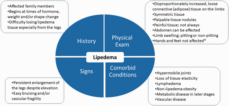
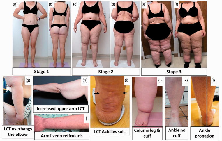
Background: Lipedema is a loose connective tissue disease predominantly in women identified by increased nodular and fibrotic adipose tissue on the buttocks, hips and limbs that develops at times of hormone, weight and shape change including puberty, pregnancy, and menopause. Lipedema tissue may be very painful and can severely impair mobility. Non-lipedema obesity, lymphedema, venous disease, and hypermobile joints are comorbidities. Lipedema tissue is difficult to reduce by diet, exercise, or bariatric surgery.

Methods: This paper is a consensus guideline on lipedema written by a US committee following the Delphi Method. Consensus statements are rated for strength using the GRADE system.

Results: Eighty-five consensus statements outline lipedema pathophysiology, and medical, surgical, vascular, and other therapeutic recommendations. Future research topics are suggested.

Conclusion: These guidelines improve the understanding of the loose connective tissue disease, lipedema, to advance our understanding towards early diagnosis, treatments, and ultimately a cure for affected individuals.

Keywords: Lipedema; chronic venous disease; hypermobility; lymphedema; standard of care.


Conflict of interest statement

Declaration of Conflicting Interests: The author(s) declared the following potential conflicts of interest with respect to the research, authorship, and/or publication of this article: KLH received research funding from Raziel Therapeutics, is on the Speaker’s Board for Tactile Medical, and has received honorarium for speaking engagements from Sigvaris and Lymphapress. MS received consulting money from Microaire. LBM received honorarium for speaking engagements for Lymphapress. LAK received honorarium for speaking engagements from Compression Guru. SMD is on the Speaker’s Bureau and Scientific Advisory Board for Tactile Medical. TFW received research funding from Raziel Therapeutics and has received honorarium for speaking engagements from Sigvaris and Tactile Medical. PCD is a consultant for PureTech Health and received grant funding from LymphaTouch. KL is on the Advisory Board for Aria Health. EI received honorarium for speaking engagements for Sigvaris. NJP receives consulting fees from the Obesity Medicine Association, has an independent contractor relationship with Medifast, and has received speaking fees from Integrity Continuing Education, Inc.

Figures

Figure 1.
Figure 1.
Diagnostic considerations for lipedema supported by expert opinion of the United States standard of care committee. *∼30% of women with lipedema can have fat tissue on the hands likely due to loss of elasticity in the tissue.
Figure 2.
Figure 2.
Stages and features of lipedema. (a) to (f): Front and back pictures of women with lipedema Stages 1 to 3. Staging references the legs, however women pictured also have arm involvement. Stage 1 skin has a smooth texture with subdermal pebble-like feel due to underlying loose connective tissue fibrosis. Lipedema Stage 2 women have more lipedema tissue than women with Stage 1 and skin dimpling due to progressed fibrotic changes and excess tissue. Palpable nodules may be more numerous and larger. Note the full Achilles sulci in pictures (d) to (f). In Lipedema Stage 2 arms, the tissue begins to hang off the arm and full arm involvement shows a more pronounced wrist cuff. Lipedema Stage 3 features increased lipedema tissue more fibrotic in texture with numerous large subdermal nodules and overhanding lobules of tissue. Patient (e) and (f) has lipedema, non-lipedema obesity and lipolymphedema. Types I to V describe the locations of lipedema tissue. Type I, lipedema tissue is present under the umbilicus and over hips and buttocks, Type II, under the umbilicus to knees (a, b), Type III, under the umbilicus to ankles (c to f), Type IV, arms (a to f) and Type V, lower legs (not shown). A tissue cuff at the ankle or wrist may be present in all stages. (g): Lipedema tissue overhangs the elbow. (h): Lipedema tissue often hangs well below the arm due to loss of elasticity and heaviness of the tissue. (i): Livedo reticularis is often a feature of lipedema. (j): Close view of tissue filling the Achilles sulci. (k): Close view of a column type lipedema leg with an obvious ankle cuff. (l): An ankle of a woman with lipedema without an ankle cuff (compare to (k)). (m): Pronation of the ankle commonly found in women with lipedema. Consent was obtained for use of all photos. LCT: loose connective tissue.
Figure 3.
Figure 3.
Nodules and thickened extracellular matrix fibers in lipedema calf loose connective tissue. (a) Example of thick fibrotic fibers (white arrowhead) connecting skin to superficial fascia (*). The abnormal fibers when palpated through the wound are firm and thick and less mobile due to fibrosis in comparison to adjacent fibers. (b) Three nodules under the skin (black arrows) that can be palpated through the skin as firm and that when removed feel firm. Notice extensive scar under the skin (white arrowheads). (c) Lipedema nodules (black arrows) intermingled amongst yellow fat obtained during modified suction lipectomy. Source: Photos courtesy of Jaime Schwartz.

References

    1. Buck DW, 2nd, Herbst KL. Lipedema: a relatively common disease with extremely common misconceptions. Plast Reconstr Surg Glob Open 2016; 4: e1043. - PMC - PubMed
    1. Felmerer G, Stylianaki A, Hägerling R, et al.. Adipose tissue hypertrophy, an aberrant biochemical profile and distinct gene expression in lipedema. J Surg Res 2020; 253: 294–303. - PubMed
    1. Al-Ghadban S, Cromer W, Allen M, et al.. Dilated blood and lymphatic microvessels, angiogenesis, increased macrophages, and adipocyte hypertrophy in lipedema thigh skin and fat tissue. J Obes 2019; 2019: 1–10. - PMC - PubMed
    1. Herbst K, Mirkovskaya L, Bharhagava A, et al.. Lipedema fat and signs and symptoms of illness, increase with advancing stage. Arch Med 2015; 7: 1–8.
    1. Allen EV, Hines EAJ. Lipedema of the legs: a syndrome characterised by fat legs and orthostatic edema. Proc Staff Meet Mayo Clin 1940; 15: 184–187.