. 2021 May 28;21(1):243.
doi: 10.1186/s12876-021-01827-0.

Selective inhibition of soluble TNF using XPro1595 relieves pain and attenuates cerulein-induced pathology in mice

Affiliations

Selective inhibition of soluble TNF using XPro1595 relieves pain and attenuates cerulein-induced pathology in mice

Rajasa Randhi et al. BMC Gastroenterol. .

Abstract

Background: Symptoms associated with acute pancreatitis can be debilitating, and treatment remains a challenge. This study aimed to investigate the efficacy of selectively inhibiting the soluble form of TNF (solTNF) using the biologic XPro1595 in a mouse model of acute pancreatitis.

Methods: Acute pancreatitis was induced in adult male C57Bl/6J mice by administering cerulein (8 injections of 50 µg/kg I.P., spaced an hour apart), with XPro1595 (10 mg/kg, S.C.) or vehicle being administered approximately 18 h after the last injection. Serum was collected 6 or 18 h after the last cerulein injection, pancreatic tissue was collected 2 and 7 days post-induction, and brain hippocampal tissue was collected at 7 days post-induction. The animal's pain level was assessed 3, 5 and 7 days post-induction.

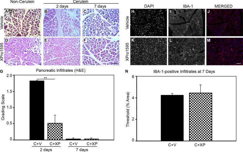
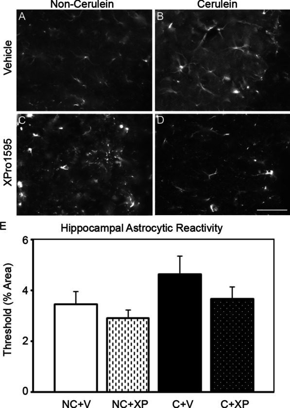
Results: The induction of acute pancreatitis promoted a strong increase in serum amylase levels, which had receded back to baseline levels by the next morning. XPro1595 treatment began after amylase levels had subsided at 18 h, and prevented pancreatic immune cell infiltration, that subsequently prevented tissue disruption and acinar cell death. These improvements in pathology were associated with a significant reduction in mechanical hypersensitivity (neuropathic pain). XPro1595 treatment also prevented an increase in hippocampal astrocyte reactivity, that may be associated with the prevention of neuropathic pain in this mouse model.

Conclusion: Overall, we observed that selectively inhibiting solTNF using XPro1595 improved the pathophysiological and neurological sequelae of cerulein-induced pancreatitis in mice, which provides support of its use in patients with pancreatitis.

Keywords: Acute pancreatitis; Cerulein; Cytokines; Inflammation; Mice; Neuropathic pain; TNF; TNFR1.


Conflict of interest statement

The authors declare that they have no competing interests.

Figures

Fig. 1
Fig. 1
Cerulein administration promotes induction of pancreatitis. Acute pancreatitis was induced by 8 intraperitoneal injections of cerulein (50 µg/kg) or vehicle to mice, spaced an hour apart each, and serum amylase concentrations were measured 6 and 18 h after the last cerulein injection. Cerulein-treated mice displayed a temporary spike in serum amylase concentration at 6 h, that had reduced back to baseline levels by the next morning (n = 3 per group). NC non-cerulein, C cerulein, **p < 0.01, ***p < 0.001
Fig. 2
Fig. 2
XPro1595 treatment attenuates cerulein-induced pancreatic inflammatory infiltrates. Photomicrographs illustrate hematoxylin and eosin stained sections of mouse pancreas in non-cerulein (a, d) and cerulein (b, c, e, f) mice, 2 days after cerulein/vehicle administration. Vehicle-treated non-cerulein mice had an absence of inflammatory infiltrates (a) (n = 5), but cerulein treatment promoted an influx of inflammatory cells within 2 days (b) (n = 3), which had resolved by 7 days (c, d) (n = 4). XPro1595 treatment significantly reduced the immune cell infiltration at 2 days (e) (n = 3), that was even further reduced by 7 days (f) (n = 4). A week following pancreatitis induction the circulating macrophage population was also minimal in cerulein-treated mice, independent of vehicle or XPro1595 treatment (n = 4 each). C cerulein, V vehicle, XP XPro1595, **p < 0.01, scale bars = 100 µm
Fig. 3
Fig. 3
Selective inhibition of solTNF in mice with acute pancreatitis prevents pancreatic tissue degradation at 7 days after induction. Photomicrographs illustrate H&E stained pancreatic sections from mice with acute pancreatitis (cerulein treated) with either vehicle—a (n = 4) or XPro1595-treatment b (n = 4), where quantification shows vehicle-treated pancreatitis mice have significant deterioration of tissue integrity (c), compared to pancreatitis mice treated with XPro1595 (n = 4), or non-pancreatitis mice (n = 3–4). In accordance, the vehicle-treated pancreatitis mice also display an exacerbated level of pancreatic acinar cell atrophy (d) that is not observed in the XPro1595 treated pancreatitis group (e, f). A week after induction of pancreatitis the intralobular ducts in the vehicle-treated group appeared to be invaded by groups of acini spreading apart (g), although this was less prominent in the XPro1595-treated group (h, i). NC non-cerulein, C cerulein, V vehicle, XP XPro1595, *p < 0.05, **p < 0.01, ***p < 0.001, scale bar in a and b = 500 µm, scalebar in d and e = 25 µm, scalebar in g and h = 500 µm
Fig. 4
Fig. 4
Soluble TNF activity reduces hindpaw mechanical hyper-sensitivity following induction of acute pancreatitis in mice. Graph shows mechanical hypersensitivity of the hindpaw when touched by von Frey filaments, as a percentage of pre-cerulein baseline data for each mouse. No differences were observed in hypersensitivity of non-pancreatitis mice between baseline and on days 3, 5 and 7, independent of treatment (n = 6–8). In contrast, vehicle-treated pancreatitis mice displayed significantly more hypersensitivity than non-pancreatitis control mice at each time point assessed during the first week (n = 9). Treating mice with XPro1595 prevented this hypersensitivity at all time-points tested (n = 9), and this group displayed significantly less hypersensitivity than the vehicle-treated pancreatitis group on the last 2 days of testing. NC non-cerulein, C cerulein, V vehicle, XP XPro1595, *p < 0.05, ***p < 0.001
Fig. 5
Fig. 5
Hippocampal GFAP expression is more pronounced in cerulein-treated mice without XPro1595 administration. CA1 hippocampal GFAP expression was semi-quantitated 7 days following induction of pancreatitis in mice. Non-pancreatitis mouse groups, independent of vehicle or XPro1595 treatment, did not display reactive astrogliosis (a, c) (n = 3–4). In contrast, the vehicle-treated pancreatitis tissue (b) had numerous reactive astrocytes and when quantitated displayed a tendency for increased GFAP expression (n = 4), compared to the non-pancreatitis groups, but was not statistically significantly different (e). XPro1595 treated pancreatitis mice prevented the induction of reactive astrocytes (d) (n = 4). NC non-cerulein, C cerulein, V vehicle, XP XPro1595, scalebar = 100 µm

Similar articles

Cited by

  • TNF and TNF receptors as therapeutic targets for rheumatic diseases and beyond.
    Siegmund D, Wajant H. Siegmund D, et al. Nat Rev Rheumatol. 2023 Sep;19(9):576-591. doi: 10.1038/s41584-023-01002-7. Epub 2023 Aug 4. Nat Rev Rheumatol. 2023. PMID: 37542139 Review.
  • Apoptotic cell death in disease-Current understanding of the NCCD 2023.
    Vitale I, Pietrocola F, Guilbaud E, Aaronson SA, Abrams JM, Adam D, Agostini M, Agostinis P, Alnemri ES, Altucci L, Amelio I, Andrews DW, Aqeilan RI, Arama E, Baehrecke EH, Balachandran S, Bano D, Barlev NA, Bartek J, Bazan NG, Becker C, Bernassola F, Bertrand MJM, Bianchi ME, Blagosklonny MV, Blander JM, Blandino G, Blomgren K, Borner C, Bortner CD, Bove P, Boya P, Brenner C, Broz P, Brunner T, Damgaard RB, Calin GA, Campanella M, Candi E, Carbone M, Carmona-Gutierrez D, Cecconi F, Chan FK, Chen GQ, Chen Q, Chen YH, Cheng EH, Chipuk JE, Cidlowski JA, Ciechanover A, Ciliberto G, Conrad M, Cubillos-Ruiz JR, Czabotar PE, D'Angiolella V, Daugaard M, Dawson TM, Dawson VL, De Maria R, De Strooper B, Debatin KM, Deberardinis RJ, Degterev A, Del Sal G, Deshmukh M, Di Virgilio F, Diederich M, Dixon SJ, Dynlacht BD, El-Deiry WS, Elrod JW, Engeland K, Fimia GM, Galassi C, Ganini C, Garcia-Saez AJ, Garg AD, Garrido C, Gavathiotis E, Gerlic M, Ghosh S, Green DR, Greene LA, Gronemeyer H, Häcker G, Hajnóczky G, Hardwick JM, Haupt Y, He S, Heery DM, Hengartner MO, Hetz C, Hildeman DA, Ichijo H, Inoue S, Jäättelä M, Janic A, Joseph B, Jost PJ, Kanneganti TD, Karin M, Kashkar H, Kaufmann T, Kelly … See abstract for full author list ➔ Vitale I, et al. Cell Death Differ. 2023 May;30(5):1097-1154. doi: 10.1038/s41418-023-01153-w. Epub 2023 Apr 26. Cell Death Differ. 2023. PMID: 37100955 Free PMC article. Review.
  • Blueprint of Collapse: Precision Biomarkers, Molecular Cascades, and the Engineered Decline of Fast-Progressing ALS.
    Șerban M, Toader C, Covache-Busuioc RA. Șerban M, et al. Int J Mol Sci. 2025 Aug 21;26(16):8072. doi: 10.3390/ijms26168072. Int J Mol Sci. 2025. PMID: 40869392 Free PMC article. Review.

References

    1. Bugiantella W, Rondelli F, Boni M, Stella P, Polistena A, Sanguinetti A, et al. Necrotizing pancreatitis: a review of the interventions. Int J Surg. 2016;28(Suppl 1):S163–S171. doi: 10.1016/j.ijsu.2015.12.038. - DOI - PubMed
    1. Malleo G, Mazzon E, Siriwardena AK, Cuzzocrea S. Role of tumor necrosis factor-alpha in acute pancreatitis: from biological basis to clinical evidence. Shock. 2007;28(2):130–140. doi: 10.1097/shk.0b013e3180487ba1. - DOI - PubMed
    1. Hughes CB, Gaber LW, Kotb M, Mohey el-Din AB, Pabst M, Gaber AO. Induction of acute pancreatitis in germ-free rats: evidence of a primary role for tumor necrosis factor-alpha. Surgery. 1995;117(2):201–5. doi: 10.1016/S0039-6060(05)80086-8. - DOI - PubMed
    1. Grewal HP, Kotb M, el Din AM, Ohman M, Salem A, Gaber L, et al. Induction of tumor necrosis factor in severe acute pancreatitis and its subsequent reduction after hepatic passage. Surgery. 1994;115(2):213–221. - PubMed
    1. Norman JG, Fink GW, Franz MG. Acute pancreatitis induces intrapancreatic tumor necrosis factor gene expression. Arch Surg. 1995;130(9):966–970. doi: 10.1001/archsurg.1995.01430090052018. - DOI - PubMed