Observation of the characteristics of the natural course of Bietti crystalline dystrophy by fundus fluorescein angiography
- PMID: 34049507
- PMCID: PMC8161580
- DOI: 10.1186/s12886-021-01999-z
Observation of the characteristics of the natural course of Bietti crystalline dystrophy by fundus fluorescein angiography
Abstract
Background: Bietti crystalline dystrophy (BCD) is an autosomal recessive genetic disorder that causes progressive vision loss. Here, 12 patients were followed up for 1-5 years with fundus fluorescein angiography (FFA) to observe BCD disease progression.
Methods: FFA images were collected for 12 patients with BCD who visited our clinic twice or more over a 5-year period. Peripheral venous blood was collected to identify the pathogenic gene related to the clinical phenotype.
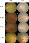
Results: We observed two types in FFA images of patients with BCD. Type 1 showed retinal pigment epithelium (RPE) atrophy in the macular area, followed by choriocapillaris atrophy and the subsequent appearance of RPE atrophy appeared at the peripheral retina. Type 2 showed RPE atrophy at the posterior pole and peripheral retina, followed by choriocapillaris atrophy around the macula and along the superior and inferior vascular arcades and the nasal side of the optic disc. The posterior and peripheral lesions of both type 1 and type 2 BCD subsequently extended to the mid-periphery; finally, all the RPEs and choriocapillaris atrophied, exposing the choroid great vessels, but type 2 macular RPE atrophy could last longer.
Conclusions: The characterization of two different types of BCD development provides a better understanding of the phenotype and the progression of the disease for a precise prognosis and prediction of pathogenesis.
Keywords: Bietti crystalline dystrophy; Disease development; Fundus fluorescein angiography.
Conflict of interest statement
The authors declare that they have no competing interests.
Figures


Similar articles
-
Multimodal Imaging Observation in Different Progressive Types of Bietti Crystalline Dystrophy.J Ophthalmol. 2022 May 24;2022:7426052. doi: 10.1155/2022/7426052. eCollection 2022. J Ophthalmol. 2022. PMID: 35655804 Free PMC article.
-
Multimodal imaging features and genetic findings in Bietti crystalline dystrophy.BMC Ophthalmol. 2020 Aug 15;20(1):331. doi: 10.1186/s12886-020-01545-3. BMC Ophthalmol. 2020. PMID: 32799831 Free PMC article.
-
Clinical and molecular findings in three Lebanese families with Bietti crystalline dystrophy: report on a novel mutation.Mol Vis. 2012;18:1182-8. Epub 2012 May 5. Mol Vis. 2012. PMID: 22605929 Free PMC article.
-
[A new approach for studying the retinal and choroidal circulation].Nippon Ganka Gakkai Zasshi. 2004 Dec;108(12):836-61; discussion 862. Nippon Ganka Gakkai Zasshi. 2004. PMID: 15656089 Review. Japanese.
-
Genetics of Bietti Crystalline Dystrophy.Asia Pac J Ophthalmol (Phila). 2016 Jul-Aug;5(4):245-52. doi: 10.1097/APO.0000000000000209. Asia Pac J Ophthalmol (Phila). 2016. PMID: 27228076 Review.
Cited by
-
Longitudinal structure-function analysis of molecularly-confirmed CYP4V2 Bietti Crystalline Dystrophy.Eye (Lond). 2024 Apr;38(5):853-862. doi: 10.1038/s41433-023-02791-7. Epub 2023 Oct 28. Eye (Lond). 2024. PMID: 37898718 Free PMC article.
-
Presumed Bietti crystalline dystrophy with optic nerve head drusen: a case report.J Med Case Rep. 2022 Nov 2;16(1):413. doi: 10.1186/s13256-022-03581-7. J Med Case Rep. 2022. PMID: 36320086 Free PMC article.
-
Multimodal Imaging Observation in Different Progressive Types of Bietti Crystalline Dystrophy.J Ophthalmol. 2022 May 24;2022:7426052. doi: 10.1155/2022/7426052. eCollection 2022. J Ophthalmol. 2022. PMID: 35655804 Free PMC article.
-
Longitudinal quantitative assessment of retinal crystalline deposits in bietti crystalline dystrophy.BMC Ophthalmol. 2025 Mar 17;25(1):139. doi: 10.1186/s12886-025-03962-8. BMC Ophthalmol. 2025. PMID: 40097952 Free PMC article.
-
Optical coherence tomography angiography in Bietti crystalline dystrophy.Eur J Ophthalmol. 2023 Nov;33(6):NP1-NP5. doi: 10.1177/11206721221143156. Epub 2022 Dec 1. Eur J Ophthalmol. 2023. PMID: 36457241 Free PMC article.
References
-
- Bietti GB. Ueber familiares Vorkommen von “Retinitis punctata albescens” (verbunden mit “Dystrophia marginaliscristallinea cornea”): Glitzern des Glaskörpers und anderen degenerativen Augenveränderungen. Klin Monatsbl Augenheilkd. 1937;99:737–56.
-
- Bietti GB. Su alcune forme atipiche o rare di degenerazione retinica (degenerazioni tappetoretiniche e quadri morbosi similari) Boll Oculist. 1937;16:1159–244.
MeSH terms
Supplementary concepts
LinkOut - more resources
Full Text Sources
Other Literature Sources

