. 2021 May;9(5):e002432.
doi: 10.1136/jitc-2021-002432.

In situ delivery of iPSC-derived dendritic cells with local radiotherapy generates systemic antitumor immunity and potentiates PD-L1 blockade in preclinical poorly immunogenic tumor models

Affiliations

In situ delivery of iPSC-derived dendritic cells with local radiotherapy generates systemic antitumor immunity and potentiates PD-L1 blockade in preclinical poorly immunogenic tumor models

Takaaki Oba et al. J Immunother Cancer. 2021 May.

Abstract

Background: Dendritic cells (DCs) are a promising therapeutic target in cancer immunotherapy given their ability to prime antigen-specific T cells, and initiate antitumor immune response. A major obstacle for DC-based immunotherapy is the difficulty to obtain a sufficient number of functional DCs. Theoretically, this limitation can be overcome by using induced pluripotent stem cells (iPSCs); however, therapeutic strategies to engage iPSC-derived DCs (iPSC-DCs) into cancer immunotherapy remain to be elucidated. Accumulating evidence showing that induction of tumor-residing DCs enhances immunomodulatory effect of radiotherapy (RT) prompted us to investigate antitumor efficacy of combining intratumoral administration of iPSC-DCs with local RT.

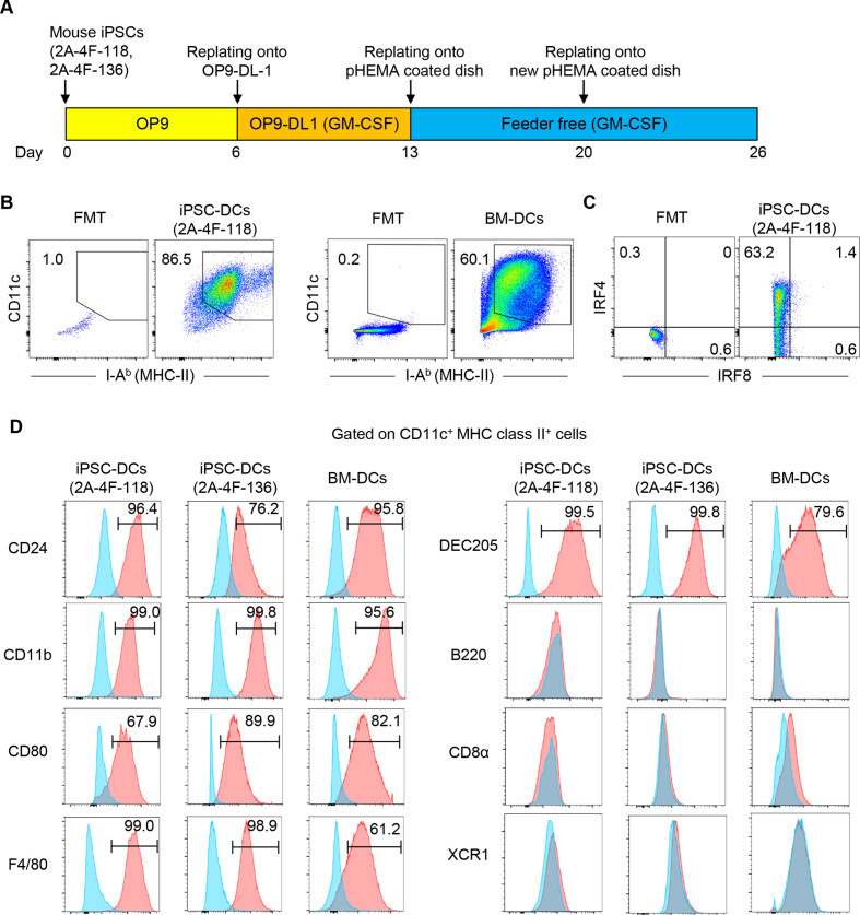
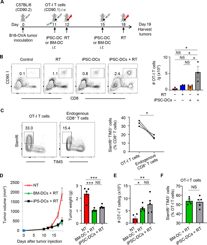
Methods: Mouse iPSCs were differentiated to iPSC-DCs on OP9 stromal cells expressing the notch ligand delta-like 1 in the presence of granulocyte macrophage colony-stimulating factor. Phenotype and the capacities of iPSC-DCs to traffic tumor-draining lymph nodes (TdLNs) and prime antigen-specific T cells were evaluated by flow cytometry and imaging flow cytometry. Antitumor efficacy of intratumoral injection of iPSC-DCs and RT was tested in syngeneic orthotopic mouse tumor models resistant to anti-PD-1 ligand 1 (PD-L1) therapy.

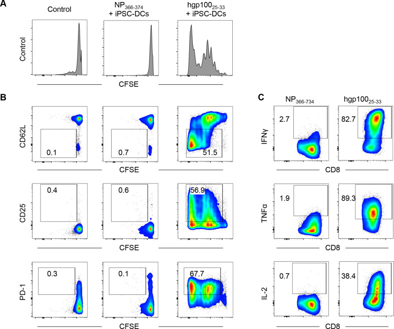
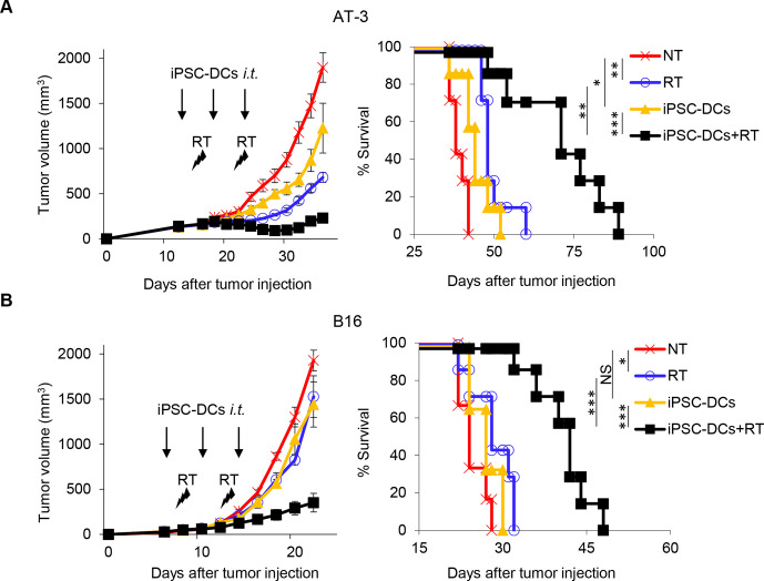
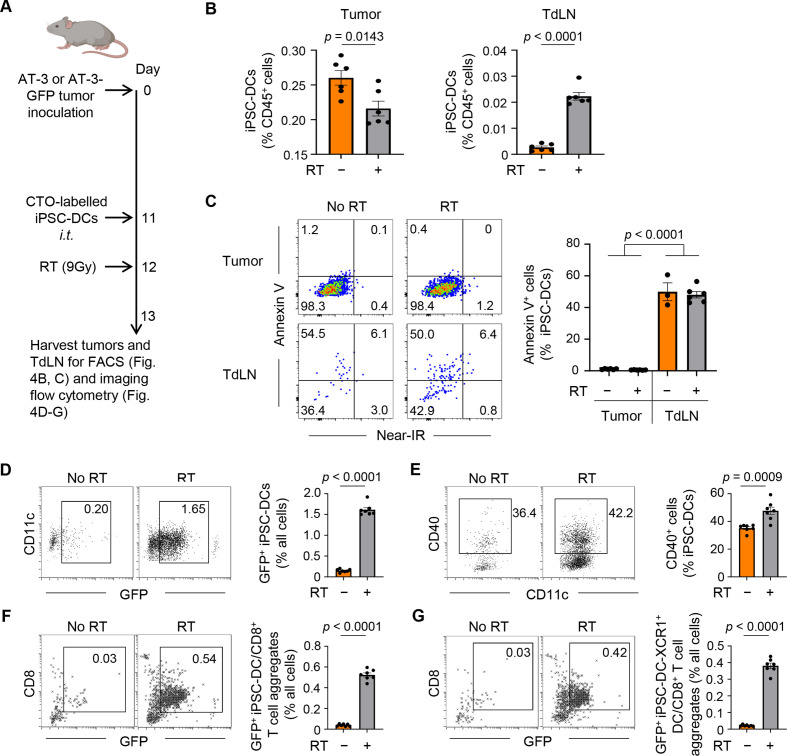
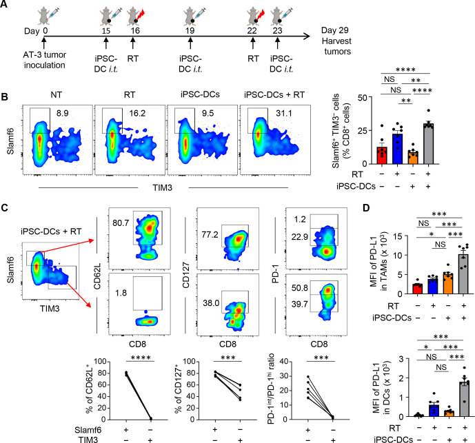
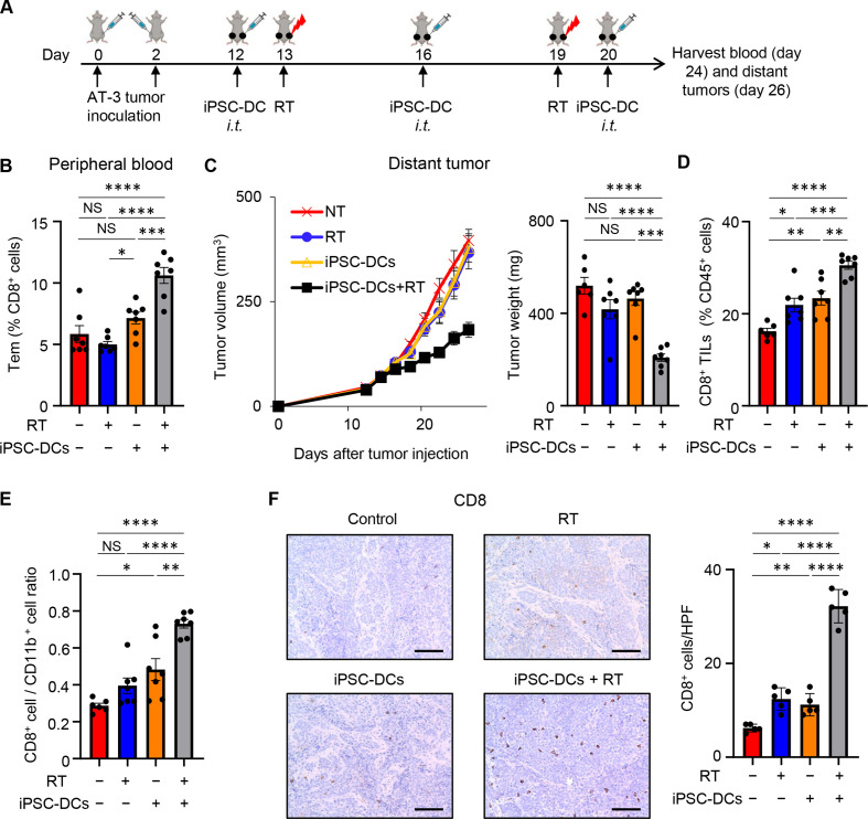
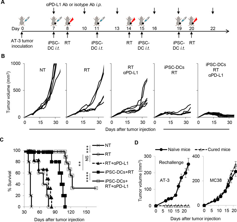
Results: Mouse iPSC-DCs phenotypically resembled conventional type 2 DCs, and had a capacity to promote activation, proliferation and effector differentiation of antigen-specific CD8+ T cells in the presence of the cognate antigen in vitro. Combination of in situ administration of iPSC-DCs and RT facilitated the priming of tumor-specific CD8+ T cells, and synergistically delayed the growth of not only the treated tumor but also the distant non-irradiated tumors. Mechanistically, RT enhanced trafficking of intratumorally injected iPSC-DCs to the TdLN, upregulated CD40 expression, and increased the frequency of DC/CD8+ T cell aggregates. Phenotypic analysis of tumor-infiltrating CD8+ T cells and myeloid cells revealed an increase of stem-like Slamf6+ TIM3- CD8+ T cells and PD-L1 expression in tumor-associated macrophages and DCs. Consequently, combined therapy rendered poorly immunogenic tumors responsive to anti-PD-L1 therapy along with the development of tumor-specific immunological memory.

Conclusions: Our findings illustrate the translational potential of iPSC-DCs, and identify the therapeutic efficacy of a combinatorial platform to engage them for overcoming resistance to anti-PD-L1 therapy in poorly immunogenic tumors.

Keywords: adaptive immunity; dendritic cells; programmed cell death 1 receptor; radioimmunotherapy; vaccination.


Conflict of interest statement

Competing interests: AO was a co-founder of Tactiva Therapeutics and receives research support from AstraZeneca and Tessaro.

Figures

Figure 1
Figure 1
Generation of dendritic cells (DCs) from mouse induced pluripotent stem cells (iPSCs). (A) Schematic illustration showing the generation of DCs from iPSCs. (B) Phenotypic analysis of iPSC-derived cells on day 26, and bone marrow-derived cells on day 10 differentiated in the presence of GM-CSF in vitro. (C) Representative flow cytometric plots showing IRF4 and IRF8 expression of CD11c+ MHC class II+ iPSC-derived DCs (iPSC-DCs). (D) Phenotypic analysis of CD11c+ MHC class II+ cells. Representative histogram showing CD24, DEC205, CD80, CD11b, B220, CD8α, XCR1 and F4/80 expressions on CD11c+ MHC class II+ 2A-4F-118 iPSC-DCs, 2A-4F-136 iPSC-DCs and bone marrow-derived DCs (BM-DCs) (red). Isotype-matched controls are shown in blue. Number denotes per cent positive cells for each marker. Data shown are representative of three independent experiments. FMT, fluorescence minus two; GM-CSF, granulocyte macrophage colony-stimulating factor; IRF4/8, interferon regulatory factor-4 and 8; MHC, Major Histocompatibility Complex; OP9-DL-1, OP9 cells expressing a notch ligand, delta-like-1; pHEMA, poly 2-hydroxyethyl methacrylate.
Figure 2
Figure 2
iPSC-derived DCs (iPSC-DCs) activate antigen-specific CD8+ T cells in vitro. (A, B) Pmel-1 T cells (CD90.1+ CD8+) were isolated from splenocytes by CD8α positive selection, labeled with CFSE, and co-cultured with iPSC-DCs in the presence of influenza nucleoprotein (NP) epitope NP366–374 or hgp10025–33 and IL-2 (60 IU/mL). Two days later, cells were harvested for flow cytometric analysis. (A) Representative histogram showing CFSE dilution in Pmel-1 T cells. (B) Representative flow cytometric plots showing CD62L, CD25, and PD-1 expression in Pmel-1 T cells. Numbers denote per cent dividing CD62L, CD25+, or PD-1+ cells. (C) Activated Pmel-1 T cells from the experiment (A, B) were co-cultured with hgp10025–33 or NP366–374 in the presence of antigen-presenting cells (splenocytes from C57BL/6 mice) for another 2 days. Representative flow cytometric plots show intracellular production of IFNγ, TNFα, and IL-2 in Pmel-1 T cells activated by iPSC-DCs. Numbers denote per cent positive cells. Data shown are representative of two independent experiments. CFSE, carboxyfluorescein succinimidyl ester; IFNγ, interferon gamma; IL-2, interleukin 2; PD-1, programmed cell death protein 1; TNFα, tumor necrosis factor alpha.
Figure 3
Figure 3
Synergistic antitumor efficacy of in situ iPSC-DC administration and local radiotherapy (RT) against poorly immunogenic tumors. (A, B) Tumor volume curves (mean) and survival curves in AT-3 (n=7) (A) and B16 (n=6–7) (B) tumor-bearing mice in different treatment as indicated. NT, non treatment. Individual tumor volume curves are shown in online supplemental figure 1. NS not significant, *p<0.05, **p<0.01, ***p<0.001 by a log-rank test. Data shown are representative of two independent experiments. Mean±SEM. iPSC-DCs, induced pluripotent stem cell-derived dendritic cells; i.t., intratumorally.
Figure 4
Figure 4
RT augments trafficking of intratumorally injected iPSC-DCs to the TdLN, upregulates CD40 expression, and increases the frequency of DC/CD8+ T cell aggregates. (A) Experimental set-up. Mice bearing AT-3 or AT-3-GFP tumors had an intratumoral iPSC-DC injection with or without local RT (9 Gy). Gating strategy for identifying fluorescently (CTO: CellTracker Orange)-labeled iPSC-DCs by flow cytometry and imaging flow cytometry is shown in online supplemental figure 4 and online supplemental figure 5, respectively. FACS, fluorescence-activated cell sorting. (B) Frequency of iPSC-DCs in the tumor and TdLN in different treatment groups as indicated (n=6). (C) Representative flow cytometric plots showing annexin V and near-IR expression of iPSC-DCs in the tumor and TdLN. The data panel shows the frequency of annexin V+ iPSC-DCs in the tumor and TdLN (n=3–6). Data with total iPSC-DCs >10 from the experiment (B) were evaluated. (D–G) Representative dot plots and frequencies of GFP+ iPSC-DCs as single cells or engaged in cell aggregates (D), CD40+ CD11c+ cells in iPSC-DCs (E), GFP+ iPSC-DC in direct contact with CD8+ T-cell (F), and GFP+ iPSC-DC-XCR1+ DC in direct contact with CD8+ T-cell (G) analyzed by imaging flow cytometry. Representative images are shown in online supplemental figure 6. Each dot represents biologically independent mice (B–G). One-way ANOVA with Tukey’s multiple comparisons (C) and two-tailed unpaired t-test (B, D–G). Mean±SEM. ANOVA, analysis of variance; GFP, green fluorescent protein; iPSC-DCs, induced pluripotent stem cell-derived dendritic cells; IR, infrared; i.t., intratumorally; RT, radiotherapy; TdLN, tumor-draining lymph node.
Figure 5
Figure 5
A combination of in situ iPSC-DC injection and RT increases stem-like progenitor exhausted CD8+ T cells and PD-L1 expression in myeloid cells in the tumor. (A) Experimental set-up. (B) Phenotypic analysis of CD8+ T cells among CD45+ cells in AT-3 tumors in different treatment groups as indicated. Numbers denote per cent Slamf6+ TIM3 cells. Representative flow cytometric plots showing expression of Slamf6 and TIM3 in CD8+ TILs. The data panel shows the frequency of Slamf6+ TIM3 cells in CD8+ TILs (n=7). (C) Phenotypic characterization of Slamf6+ TIM3 and Slamf6 TIM3+ CD8+ TILs (n=6). (D) PD-L1 expression (MFI: median fluorescence intensity) of Ly6c CD11c+ class II+ F4/80hi CD24− tumor-associated macrophages (TAMs) (upper) and Ly6c CD11c+ class II+ F4/80lo CD24+ DCs (lower) in AT-3 tumors (n=7). Gating strategy for identifying TAMs and DCs is shown in online supplemental figure 3. NS not significant, *p<0.05, **p<0.01, ***p<0.001, ****p<0.0001 by one-way ANOVA with Tukey’s multiple comparisons (B, D) and two-tailed paired t-test (C). Each dot represents biologically independent mice (B, D). Data shown are representative of two independent experiments. Mean±SEM. ANOVA, analysis of variance; iPSC-DC, induced pluripotent stem cell-derived dendritic cells; i.t., intratumorally; PD-1, programmed cell death protein 1; PD-L1, PD-1 ligand 1; RT, radiotherapy; TILs, tumor-infiltrating lymphocytes; TIM3, T cell immunoglobulin and mucin domain-containing protein 3.
Figure 6
Figure 6
In situ injection of iPSC-DCs with local RT increases antigen-specific CD8+ T cell infiltrates in the tumor. (A) Experimental set-up. (B) Representative flow cytometric plots showing OT-I T cells (CD90.1+ CD8+) in CD45+ cells in tumors. Data panels show numbers (/g) (tumor) of OT-I T cells (n=3). (C) Representative flow cytometric plots showing expression of Slamf6 and TIM3 in OT-I (CD90.1+) and endogenous (CD90.2+) CD8+ TILs. Numbers denote per cent Slamf6+ TIM3 T cells. (D) Tumor growth curves (mean) (left) and tumor weight (right) of B16-OVA tumor-bearing mice in different treatment as indicated. (n=5). (E) Numbers (/g) (tumor) of OT-I T cells (n=5). Each dot represents biologically independent mice (B, D–F). NS not significant, *p<0.05, **p<0.01, ***p<0.001, one-way ANOVA with Tukey’s multiple comparisons (B, D, E) and two-tailed paired (C) and unpaired (F) t-test. Mean±SEM. ANOVA, analysis of variance; BM-DCs, bone marrow-derived dendritic cells; iPSC-DC, induced pluripotent stem cell-derived dendritic cells; i.t., intratumorally; OVA, ovalbumin; RT, radiotherapy; TILs, tumor-infiltrating lymphocytes; TIM3, T cell immunoglobulin and mucin domain-containing protein 3.
Figure 7
Figure 7
Combining in situ administration of iPSC-DC with RT attenuates growth of distant untreated tumors.(A) Experimental set-up. (B) Frequency of peripheral blood effector memory CD44+ CD62L CD8+ T cells (Tem) in different treatment groups as indicated (n=6–7). (C) Tumor growth curves (mean) (left) and tumor weight (right) of distant untreated tumors in bilateral AT-3 tumor-bearing mice in different treatment as indicated (n=6–7). Individual tumor volume curves are shown in online supplemental figure 8. (D, E). Frequency of CD8+ T cells among CD45+ cells (D) and CD8+/CD11b+ cell ratio (E) in untreated distant AT-3 tumors in different treatment groups as indicated (n=6–7). (F) Representative images of immunohistochemistry for CD8 in untreated distant AT-3 tumors. Scale bars, 100 µm. Data panels show mean numbers of CD8+ cells per each high-power field (HPF) within five different areas. Each dot represents biologically independent mice (B–E). NS not significant, *p<0.05, **p<0.01, ***p<0.001, ****p<0.0001 by one-way ANOVA with Tukey’s multiple comparisons. Data shown are representative of two independent experiments. Mean±SEM. ANOVA, analysis of variance; iPSC-DC, induced pluripotent stem cell-derived dendritic cells; i.t., intratumorally; RT, radiotherapy; TILs, tumor-infiltrating lymphocytes.
Figure 8
Figure 8
Combined therapy of in situ iPSC-DC injection and RT renders tumors responsive to anti-PD-L1 therapy, has potential to eradicate poorly T cell-inflamed tumors, and establishes immunological memory. (A) Experimental set-up. Mice bearing AT-3 tumors in the left fourth mammary gland were treated with intratumoral iPSC-DC injections, RT, and anti-PD-L1 antibody (αPD-L1 Ab) or isotype Ab. (B) Tumor growth curves (individual) in AT-3 tumor-bearing mice in different treatment groups as indicated (n=5–9). (C) Survival curves in AT-3 tumor-bearing mice in different treatment as indicated. (D) Naïve mice and surviving mice from the experiment (B, C) were rechallenged with AT-3 and MC38 in the right flank at day 123 (AT-3) and on back at day 127 (B16), respectively. NS not significant, **p<0.01, ***p<0.001, ****p=0.0001 by a log-rank test (C). Mean±SEM. Data shown are representative of two independent experiments. i.p., intraperitoneally; iPSC-DC, induced pluripotent stem cell-derived dendritic cells; i.t., intratumorally; PD-L1, PD-1 ligand 1; RT, radiotherapy.

Similar articles

Cited by

References

    1. Schuler G, Schuler-Thurner B, Steinman RM. The use of dendritic cells in cancer immunotherapy. Curr Opin Immunol 2003;15:138–47. 10.1016/s0952-7915(03)00015-3 - DOI - PubMed
    1. Palucka K, Banchereau J. Cancer immunotherapy via dendritic cells. Nat Rev Cancer 2012;12:265–77. 10.1038/nrc3258 - DOI - PMC - PubMed
    1. Wculek SK, Cueto FJ, Mujal AM, et al. . Dendritic cells in cancer immunology and immunotherapy. Nat Rev Immunol 2020;20:7–24. 10.1038/s41577-019-0210-z - DOI - PubMed
    1. Anguille S, Smits EL, Lion E, et al. . Clinical use of dendritic cells for cancer therapy. Lancet Oncol 2014;15:e257–67. 10.1016/S1470-2045(13)70585-0 - DOI - PubMed
    1. Saxena M, Balan S, Roudko V, et al. . Towards superior dendritic-cell vaccines for cancer therapy. Nat Biomed Eng 2018;2:341–6. 10.1038/s41551-018-0250-x - DOI - PMC - PubMed

Publication types

MeSH terms