. 2021 May 28;6(1):29.
doi: 10.1038/s41536-021-00139-x.

Notch-Wnt signal crosstalk regulates proliferation and differentiation of osteoprogenitor cells during intramembranous bone healing

Affiliations

Notch-Wnt signal crosstalk regulates proliferation and differentiation of osteoprogenitor cells during intramembranous bone healing

S Lee et al. NPJ Regen Med. .

Abstract

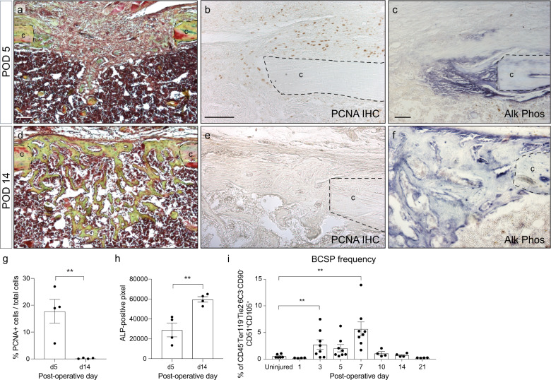
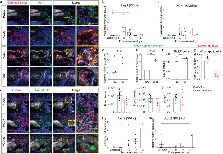
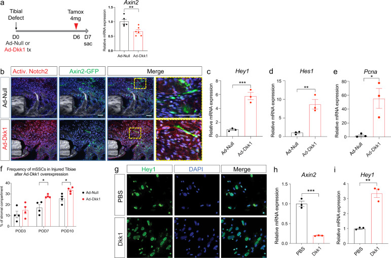
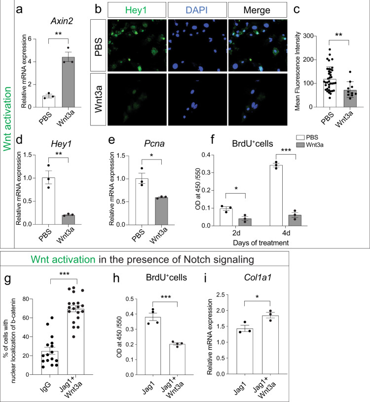
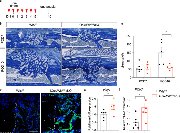
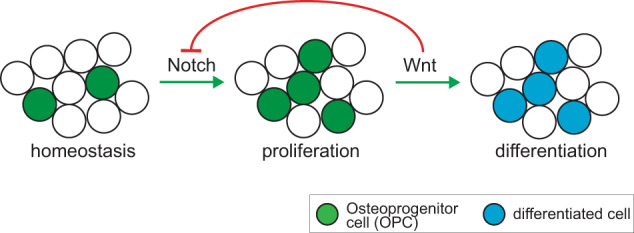
Adult bone regeneration is orchestrated by the precise actions of osteoprogenitor cells (OPCs). However, the mechanisms by which OPC proliferation and differentiation are linked and thereby regulated are yet to be defined. Here, we present evidence that during intramembranous bone formation OPC proliferation is controlled by Notch signaling, while differentiation is initiated by activation of canonical Wnt signaling. The temporospatial separation of Notch and Wnt signal activation during the early stages of bone regeneration suggests crosstalk between the two pathways. In vitro and in vivo manipulation of the two essential pathways demonstrate that Wnt activation leads to initiation of osteogenic differentiation and at the same time inhibits Notch signaling, which results in termination of the proliferative phase. Here, we establish canonical Wnt signaling as a key regulator that facilitates the crosstalk between OPC proliferation and differentiation during intramembranous, primary bone healing.


Conflict of interest statement

The authors declare no competing interests.

Figures

Fig. 1
Fig. 1. Temporal separation of proliferation and differentiation during tibial defect healing.
a Histological section of tibial mono-cortical defects 5 days after injury, stained with Movat’s Pentachrome. The defect site is filled with soft tissue. b PCNA IHC shows active proliferation in the periosteum and defect site at this early time point. c Alkaline phosphatase staining demonstrates only a small region of osteogenic differentiation at the cortical edge. d After 14 days, the defects site is filled with woven bone, stained yellow-green with Pentachrome. e PCNA staining reveals absence of proliferation within the injury. f Alkaline phosphatase staining indicating osteogenic differentiation throughout the injury site. g Quantification of PCNA-positive cells in the injury site at POD 5 and 14. h Quantification of alkaline phosphatase staining at POD 5 and 14. i Frequency of bone-cartilage-stromal progenitor cells (BCSPs) was analyzed over the time course of fracture healing by flow cytometry using the following BCSP markers: CD45 Ter119 Tie2 CD51+ CD90 6C3 CD105+. Scale bar = 100 μm. ALP alkaline phosphatase, BCSP bone-cartilage-stromal progenitor cells, c cortical bone, PCNA proliferating cell nuclear antigen, POD postoperative day. **p < 0.01. Data were represented as mean ± s.e.m.
Fig. 2
Fig. 2. Notch signaling dominates the proliferative phase while canonical Wnt signaling governs the differentiation phase.
a Temporospatial expression profile of Notch downstream target Hey1 by immunostaning and co-localization with osterix-positive cells. To label osterix-positive cells, iOsx/tdTom mice were pulsed by tamoxifen injection (1 mg/day/mouse) twice at 1 day before surgery and 1 day after surgery. b, c Temporal Notch downstream target gene expression during the early injury response (uninjured, POD 1–10) in SSCs and BCSPs (n = 6–9). d, e Notch activation using Jagged1-coated tissue culture plates resulted in upregulation of the Notch target genes Hey1 and Hes1. f In response to Notch activation, proliferation increased, shown by BrdU quantification. g The number of tdTomato-positive (osx-positive OPCs) cells decreased in the POD 3 injury site after Notch inhibition in iOSX/tdTom/Rbpjfl/fl mice. Tamoxifen (1 mg/day/mouse) was injected from day −1 until day 2. h Bone volume, i tissue volume, and j bone volume/total volume, at postoperative day 10 using microCT histomorphometry demonstrates a smaller bone volume in the regenerate in the iOSX/tdTom/Rbpjfl/fl mice (tamoxifen (1 mg/day/mouse daily) from day −1 to day 6). k Spatial expression of Wnt responsiveness within osx-positive (IF) OPCs using iAxin2/GFP reporter mice (Tamoxifen administration: 1 day before euthanasia). Scale bar = 50 μm. l, m Axin2 gene expression SSCs and BCSPs in uninjured and POD 7–21 days (n = 4). BCSP bone-cartilage-stromal progenitor cells, Jag1 Jagged1, POD postoperative day, SSC skeletal stem cell. *p < 0.05, **p < 0.01, ***p < 0.001. Data were represented as mean ± s.e.m.
Fig. 3
Fig. 3. Wnt inhibition sustains Notch activation and lengthens the proliferative phase.
a Ad-Null (control) or Ad-Dkk1 (Wnt antagonist) was administered around tibial defect sites of iAxin2/GFP reporter mice and tamoxifen was injected at POD 6 (24 h before euthanasia) (n = 5–6). Axin2 expression levels were significantly decreased at day 3 after Ad-Dkk1 administration, confirming Wnt inhibition. b Immunofluorescence staining against NICD2 (activated Notch2 intracellular domain) reveals near absent Notch activation in the control injury, while the majority of cells in the injury site treated with Ad-Dkk1 showed NICD2 staining. iAxin2/GFP IF confirmed successful Wnt inhibition. n = 3. Scale bar = 50 μm. c, d Notch target gene expression and e proliferative cell nuclear antigen (Pcna) gene expression from POD7 callus reveal activated Notch signaling in Ad-Dkk1 treated injuries with increased proliferation (n = 3). f FACS analysis of SSCs in the fracture callus of Ad-Null and Ad-Dkk1 treated animals at POD 3, 7, and 10 (n = 4) showing increase of SSC number during the proliferative phase after Wnt inhibition. g Primary bone marrow derived stromal cells were treated with PBS (control) or recombinant Dkk1 protein for 48 h in vitro. Hey1 IF shows an increased number of Notch activated cells after Dkk1 treatment. Experiments were performed in triplicate and repeated at least twice. h Axin2 expression is decreased after Dkk1 treatment. i Resulting in increased Hey1 gene expression (n = 3). PBS phosphate buffered saline. *p < 0.05, **p < 0.01, ***p < 0.001. Data were represented as mean ± s.e.m.
Fig. 4
Fig. 4. Wnt signal activation suppresses Notch and its associated proliferative effect.
a Axin2 gene expression is increased in progenitor cells treated with a recombinant protein Wnt3a (100 ng/ml) for 48 h in vitro. b Hey1 immunofluorescence, c quantification of Hey1 IF, and d gene expression of bone marrow stromal cells treated with PBS (control) or Wnt3a in vitro demonstrating Notch inhibition. e, f Decreased proliferation of bone marrow stromal cells after Wnt3a treatment shown by Pcna expression (e) and BrdU assay at 2 and 4 days in vitro (f). gi OPCs were treated in vitro with Wnt3a in presence of Notch signaling (Jag1-coated plates) and analyzed for nuclear localization of β-catenin (g), BrdU-positive proliferating cells (h), and osteogenic differentiation marker Col1a1 (i). *p < 0.05, **p < 0.01, ***p < 0.001. Experiments were performed in triplicate and repeated at least twice. Data were represented as mean ± s.e.m.
Fig. 5
Fig. 5. Wnt deletion in osx-lineage cells prolongs Notch responsiveness and associated proliferation.
a Experimental scheme using iOsx/Wlsfl/fl cKO mice. Red arrow head indicates tamoxifen i.p. injection (1 mg/day), gray arrow denotes day of tibial defect surgery and day of euthanasia. b Aniline Blue staining of tibial defect sites at POD7 and POD10 in control and iOsx/Wlsfl/fl cKO mice. c Histomorphometry of bone volume in callus of control and iOsx/Wlsfl/fl cKO mice. d Increased and prolonged Notch responsiveness in iOsx/Wlsfl/fl cKO mice shown by IF against Hey1 and e Hey1 gene expression. f PCNA expression revealed increased proliferation in the injury sites of iOsx/Wlsfl/fl cKO mice. Scale bar = 50 μm. n = 6. d Pcna expression at day 10 indicates a prolonged proliferative phase up to day 10 in the presence of decreased Wnt signaling. *p < 0.05. c cortical bone. Data were represented as mean ± s.e.m.
Fig. 6
Fig. 6. Graphical summary.
After skeletal injury, Notch signaling becomes active and initiates a proliferative response. Days later, canonical Wnt signaling is activated in the injury site, leading to active suppression of Notch signaling and its associated pro-proliferative effect. Wnt signaling then induces differentiation.

References

    1. Zura R, et al. Epidemiology of fracture nonunion in 18 human bones. JAMA Surg. 2016;151:e162775. doi: 10.1001/jamasurg.2016.2775. - DOI - PubMed
    1. Steinert AF, Rackwitz L, Gilbert F, Nöth U, Tuan RS. Concise review: the clinical application of mesenchymal stem cells for musculoskeletal regeneration: current status and perspectives. Stem Cells Transl. Med. 2012;1:237–247. doi: 10.5966/sctm.2011-0036. - DOI - PMC - PubMed
    1. Zhou BO, Yue R, Murphy MM, Peyer JG, Morrison SJ. Leptin-receptor-expressing mesenchymal stromal cells represent the main source of bone formed by adult bone marrow. Cell Stem Cell. 2014;15:154–168. doi: 10.1016/j.stem.2014.06.008. - DOI - PMC - PubMed
    1. Josephson AM, et al. Age-related inflammation triggers skeletal stem/progenitor cell dysfunction. Proc. Natl Acad. Sci. USA. 2019;116:6995–7004. doi: 10.1073/pnas.1810692116. - DOI - PMC - PubMed
    1. Kopan R, Ilagan MX. The canonical Notch signaling pathway: unfolding the activation mechanism. Cell. 2009;137:216–233. doi: 10.1016/j.cell.2009.03.045. - DOI - PMC - PubMed